Skip to main page content
U.S. flag

An official website of the United States government

Dot gov

The . gov means it’s official. Federal government websites often end in . gov or . mil. Before sharing sensitive information, make sure you’re on a federal government site VSports app下载. .

Https

The site is secure V体育官网. The https:// ensures that you are connecting to the official website and that any information you provide is encrypted and transmitted securely. .

. 2009 Mar;114(2):263-75.
doi: 10.1007/s10549-008-0011-8. Epub 2008 Apr 14.

V体育平台登录 - Activation of ErbB3, EGFR and Erk is essential for growth of human breast cancer cell lines with acquired resistance to fulvestrant

Affiliations

Activation of ErbB3, EGFR and Erk is essential for growth of human breast cancer cell lines with acquired resistance to fulvestrant

Thomas Frogne et al. Breast Cancer Res Treat. 2009 Mar.

Abstract (VSports)

Seven fulvestrant resistant cell lines derived from the estrogen receptor alpha positive MCF-7 human breast cancer cell line were used to investigate the importance of epidermal growth factor receptor (ErbB1-4) signaling. We found an increase in mRNA expression of EGFR and the ErbB3/ErbB4 ligand heregulin2 (hrg2) and a decrease of ErbB4 in all resistant cell lines. Western analyses confirmed the upregulation of EGFR and hrg2 and the downregulation of ErbB4. Elevated activation of EGFR and ErbB3 was seen in all resistant cell lines and the ErbB3 activation occurred by an autocrine mechanism. ErbB4 activation was observed only in the parental MCF-7 cells. The downstream kinases pAkt and pErk were increased in five of seven and in all seven resistant cell lines, respectively. Treatment with the EGFR inhibitor gefitinib preferentially inhibited growth and reduced the S phase fraction in the resistant cell lines concomitant with inhibition of Erk and unaltered Akt activation VSports手机版. In concert, inhibition of Erk with U0126 preferentially reduced growth of resistant cell lines. Treatment with ErbB3 neutralizing antibodies inhibited ErbB3 activation and resulted in a modest but statistically significant growth inhibition of two resistant cell lines. These data indicate that ligand activated ErbB3 and EGFR, and Erk signaling play important roles in fulvestrant resistant cell growth. Furthermore, the decreased level of ErbB4 in resistant cells may facilitate heterodimerization of ErbB3 with EGFR and ErbB2. Our data support that a concerted action against EGFR, ErbB2 and ErbB3 may be required to obtain complete growth suppression of fulvestrant resistant cells. .


Figures
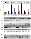
Fig. 1
Fig. 1
ErbB receptor expression and activation. (a) Real-time RT-PCR quantification of EGFR, ErbB2, ErbB3 and ErbB4 mRNA levels in MCF-7 and seven fulvestrant resistant cell lines grown in standard growth medium. As the data are presented relative to MCF-7, four independent measurements were performed on this cell line and standard deviations are shown. For the resistant cell lines, two independent determinations were done and a representative result is show. (b) Western blots showing total levels of EGFR, ErbB2, ErbB3 and ErbB4 and phosphorylated levels of ErbB2 and ErbB3 from MCF-7 and the resistant cell lines grown in their standard growth medium. Hsp70 serves as loading control. Two independent experiments were performed and similar results were obtained. (c) Immunoprecipitation of ErbB4 from MCF-7 and resistant cell lines grown in their standard growth medium. The IP membrane was analyzed for phospho-tyrosine, stripped and reproped for total ErbB4 protein. Negative control (NC) was done on MCF-7 cell lysate using total IgG from non immunized rabbits. Two independent experiments were performed and similar results were obtained
Fig. 2
Fig. 2
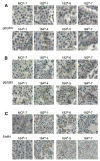
Immunohistochemical determination of pEGFR, pErbB3 and total ErbB4. Immunohistochemical staining showing (a) pEGFR; (b) pErbB3 and (c) total amount of ErbB4 in MCF-7 and resistant cell lines grown in their standard growth medium. All stainings were done simultaneously using sections from a paraffin block with all cell lines present as 2 mm cores from paraffin-embedded cell pellets. The pictures are representative of two independent experiments
Fig. 3
Fig. 3
Activation of the kinases Erk, Akt, RSK and GSK3. Western blots showing total and phosphorylated levels of the kinases Erk and Akt and their substrates RSK and GSK3, respectively. Total proteins were extracted from MCF-7 and the resistant cell lines grown in their standard growth medium. Total levels of Akt and Erk serve as loading control. Two independent experiments were performed and comparable results obtained
Fig. 4
Fig. 4
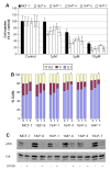
Effect of gefitinib on growth and cell cycle distribution and on ErbB2, ErbB3, Erk and Akt phosphorylation. (a) Cell numbers of MCF-7 and five resistant cell lines were determined after five days of gefitinib treatment. Cell number is expressed relative to its own vehicle treated control. Between three and six independent experiments were performed with reproducible results and a representative experiment is shown. The error bars are standard deviations. Statistical comparison of MCF-7 and the resistant cell lines was done for cultures receiving treatment with the same gefitinib concentration, and statistical significances (P < 0.05) are denoted by *. (b) Cell cycle distribution was assessed after 48 hours of treatment with gefitinib using FACS analysis. Each bar shows the distribution of the three cell cycle phases in response to treatment with 1 or 5 μM gefitinib for 48 h. The figure is showing a representative result and at least three independent experiments with reproducible results were performed. Statistically significant differences between MCF-7 and the resistant cell lines receiving the same treatment are denoted by *. (c) Western blots showing phosphorylated levels of ErbB2 and total and phosphorylated levels of ErbB3 in MCF-7 and five resistant cell lines. Hsp70 is loading control. All cell lines were grown in their standard growth medium for 3 or 4 days followed by incubation with vehicle or gefitinib for 24 h. It should be mentioned that standard growth medium for the resistant cell lines includes 100 nM fulvestrant. Three independent experiments were performed and equivalent results were observed. (d) Western blots showing total and phosphorylated levels of Erk and Akt protein in MCF-7 and the five resistant cell lines grown in their standard growth medium and incubated with 1 or 10 μM gefitinib for 24 h. Three independent experiments were performed and a representative result is shown
Fig. 5
Fig. 5
Effect of the Erk inhibitor U0126 on cell growth, cell cycle distribution and Erk activation. (a) Growth analysis of MCF-7 and five fulvestrant resistant cell lines. Each bar represents mean cell number from triplicate wells in response to treatment for five days with increasing concentrations of U0126. At least three independent experiments were performed, each in triplicate. A representative experiment with mean and SD is shown. Statistical comparison of MCF-7 and the resistant cell lines was done for cultures receiving treatment with the same U0126 concentration, and statistical significances (P < 0.05) are denoted by *. (b) FACS analysis of cell cycle distribution after 48 h of treatment with 1 or 5 μM U0126. Each bar shows the distribution of the three cell cycle phases and a representative result of at least three independent experiments is shown. Statistically significant differences between MCF-7 cells and the resistant cell lines receiving the same U0126 treatment are denoted by *. (c) Western blot analysis of lysates from MCF-7 and five fulvestrant resistant cell lines, showing pErk and Erk in response to treatment with 10 μM U0126 for 24 h
Fig. 6
Fig. 6
Effect of the ErbB3 neutralizing antibody Ab5 on ErbB3, Akt and Erk activation. Western blots of MCF-7 and 164R-5 cells measuring total and phosphorylated levels of ErbB3, Akt and Erk in response to the ErbB3 neutralizing antibody Ab5 either with or without the addition of hrg1β or GST-NRG2β (hrg2β). The cells were grown in standard growth medium incubated with Ab5 for 1 h followed by addition of vehicle, hrg1β or hrg2β for 15 min. Hsp70 is loading control. Two independent experiments were performed and similar results were obtained
Fig. 7
Fig. 7
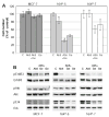
Effect of Ab5 and/or gefitinib on cell growth and ErbB3, Erk and Akt activity. (a) Growth effect of treatment with Ab5 and/or gefitinib in MCF-7, 164R-5 and 164R-7 cells. The cells were treated with vehicle (C) 10 μg/ml Ab5 (Ab5), 5 μM gefitinib (Ge) or the combination of Ab5 and gefitinib (Ab5+Ge). Cell number was determined after five days of treatment and each bar represents mean cell number from triplicate wells and standard deviations are shown. Four independent experiments were performed. Statistical analysis of the difference between control and Ab5 treatment and between gefitinib and the combination of gefitinib and Ab5 was performed and significance is denoted by *. (b) Western blots showing total and phosphorylated levels of ErbB3, Akt and Erk in MCF-7, 164R-5 and 164R-7 cells after incubation for 24 h with the indicated concentrations of Ab5 and/or gefitinib. Two independent experiments were performed and comparable results observed
Fig. 8
Fig. 8
ErbB ligand expression in MCF-7 and resistant cell lines. (a) qPCR measuring mRNA levels of the ErbB3/ErbB4 ligands hrg2α and hrg2β and (b) the EGFR/ErbB4 ligands, TGFα, HB-EGF, betacellulin, amphiregulin and epiregulin. RNA was extracted from MCF-7 and seven fulvestrant resistant cell lines grown in standard growth medium. Four independent measurements were performed on the MCF-7 cell line and standard deviations are shown. For the resistant cell lines, two independent determinations were done and similar result were observed. (c) Western blots showing hrg2α and hrg2β expression in lysates from MCF-7 and resistant cell lines grown in standard growth medium. Hsp70 is loading control. Two experiments were performed and similar results obtained. Also, three additional analyses of MCF-7, 164R-5 and 164R-7 confirmed the presented results for these cell lines. (d) Western blot showing total and phosphorylated ErbB3 in lysates from MCF-7 cells treated for 15 min. with concentrated conditioned medium from MCF-7, 164R-5 or 164R-7. As negative control (designated C) we used our regular growth medium (incl. serum), which had been concentrated in parallel to the conditioned media

References

    1. Osborne CK. Tamoxifen in the treatment of breast cancer. N Engl J Med. 1999;339:1609–1618. - PubMed
    1. Brünner N, Frandsen TL, Holst-Hansen C, et al. MCF7/LCC2 A 4-hydroxytamoxifen resistant human breast cancer variant that retains sensitivity to the steroidal antiestrogen ICI 182,780. Cancer Res. 1993;53:3229–3232. - PubMed
    1. Lykkesfeldt AE, Madsen MW, Briand P. Altered expression of estrogen-regulated genes in a tamoxifen-resistant and ICI 164,384 and ICI 182,780 sensitive human breast cancer cell line, MCF-7/TAMR-1. Cancer Res. 1994;54:1587–1595. - PubMed (VSports手机版)
    1. Howell A, DeFriend D, Robertson J, et al. Response to a specific antioestrogen (ICI 182780) in tamoxifen-resistant breast cancer. Lancet. 1995;345:29–30. - PubMed (VSports手机版)
    1. Howell A. Pure oestrogen antagonists for the treatment of advanced breast cancer. Endocr Relat Cancer. 2006;13:689–706. - PubMed

Publication types

MeSH terms

VSports在线直播 - Grants and funding