Skip to main page content
U.S. flag

An official website of the United States government

Dot gov

The . gov means it’s official. Federal government websites often end in VSports app下载. gov or . mil. Before sharing sensitive information, make sure you’re on a federal government site. .

Https

The site is secure V体育官网. The https:// ensures that you are connecting to the official website and that any information you provide is encrypted and transmitted securely. .

. 2008 May;172(5):1381-90.
doi: 10.2353/ajpath.2008.070988. Epub 2008 Apr 10.

Molecular characterization of human breast tumor vascular cells (V体育安卓版)

Affiliations

Molecular characterization of human breast tumor vascular cells (V体育ios版)

Rajendra Bhati et al. Am J Pathol. 2008 May.

Abstract

A detailed understanding of the assortment of genes that are expressed in breast tumor vessels is needed to facilitate the development of novel, molecularly targeted anti-angiogenic agents for breast cancer therapies. Rapid immunohistochemistry using factor VIII-related antibodies was performed on sections of frozen human luminal-A breast tumors (n = 5) and normal breast (n = 5), followed by laser capture microdissection of vascular cells. RNA was extracted and amplified, and fluorescently labeled cDNA was synthesized and hybridized to 44,000-element long-oligonucleotide DNA microarrays. Statistical analysis of microarray was used to compare differences in gene expression between tumor and normal vascular cells, and Expression Analysis Systematic Explorer was used to determine enrichment of gene ontology categories. Protein expression of select genes was confirmed using immunohistochemistry. Of the 1176 genes that were differentially expressed between tumor and normal vascular cells, 55 had a greater than fourfold increase in expression level VSports手机版. The extracellular matrix gene ontology category was increased while the ribosome gene ontology category was decreased. Fibroblast activation protein, secreted frizzled-related protein 2, Janus kinase 3, and neutral sphingomyelinase 2 proteins localized to breast tumor endothelium as assessed by immunohistochemistry, showing significantly greater staining compared with normal tissue. These tumor endothelial marker proteins also exhibited increased expression in breast tumor vessels compared with that in normal tissues. Therefore, these genetic markers may serve as potential targets for the development of angiogenesis inhibitors. .


Figures

Figure 1
Figure 1
LCM of human breast vascular cells. Rapid IHC for factor VIII-related antigen was followed by LCM of vascular cells. Shown are representative breast tissue specimens before and after LCM along with the collected sample containing vessels (400×).
Figure 2
Figure 2
RNA integrity analyses. High-quality RNA isolated from breast tumor vessel cells. RT-PCR primers for genes of low and high abundance levels were used on cDNA from Whole Mount, which refers to a frozen section of the whole tumor before microdissection, and LCM, which refers to the sample of vessels microdissected from a frozen section of a human breast tumor. Lane 1, DNA ladder; lane 2, 3′ end of the low expressed ADP ribosylation factor I gene (ARF F1) from the Whole Mount (239 bp); lane 3, 5′ end of ARF F1 from the Whole Mount (336 bp); lane 4, 3′ end of the housekeeping gene GAPDH from the Whole Mount (540 bp); lane 5, 5′ end of GAPDH from the Whole Mount (887 bp); lane 6, 3′ end of ARF F1 from the microdissected vessel cells; lane 7, 5′ end of ARF F1 from the microdissected vessel cells; lane 8, 3′ end of GAPDH from the microdissected vessel cells; and lane 9, 5′ end of GAPDH from the microdissected vessel cells.
Figure 3
Figure 3
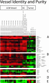
Gene expression analysis confirming vascular identity. The 44,000-feature long-oligo arrays were performed with RNA from vessel cells microdissected from five human luminal A breast tumors and five normal breast tissue samples from reduction mammoplasty, and compared to RNA from endothelial cells and breast tumor-derived cell lines cultured in vitro. Arrays for LCM vessel cells, endothelial cell lines, and breast tumor-derived cell lines were ordered from left to right. The arrays for LCM vessels were performed in duplicate, thus the designation technical replicate (techrep). Arrays from endothelial cells cultured in vitro are labeled: Dermal-microvascular-endothelial-cell, Umbilical-vein-endothelial-cell, Umbilical-vein-endothelial-cells, Aortic-smooth-muscle-cell. Arrays from breast tumor-derived cell lines in vitro are labeled: T47D-1, T47D-2, MCF7, MDA-MB-365, MDA-MB-453, HCC1937-1, and HCC1937-2. The data for different gene sets were identified, and clustered within each relevant category, in descending order: A, endothelial genes, B, TEMs, C, hematopoietic genes, D, pericyte genes, and E, epithelial genes.
Figure 4
Figure 4
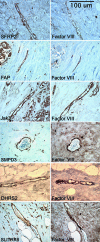
Confirmation of vascular origin of vascular marker genes. Parrafin-embedded human breast tumors were stained with antibodies to factor VIII-related antigen to identify vessels, and on the next section, an antibody to SFRP2, FAP, JAK3, SMPD3, DHRS2, or SLITRK6. Photographs were taken at ×600 magnification. Negative controls were performed with each experiment and showed no background staining (data not shown).
Figure 5
Figure 5
IHC with antibodies to FAP, SFRP2, and JAK3 on paraffin-embedded breast tumors and normal breast tissue. A high Angiogenesis Score refers to an intensity score of 3 (moderate to high staining) and percentage of enothelium staining >75%. A high Intensity Score refers to an intensity score of 3 (moderate to high staining). A: FAP had significantly higher Angiogenesis Score in luminal A, Her2/neu, and basal tumors compared to control (*P = 0.04, **P = 0.03, ***P = 0.03). B: SFRP2 had significantly higher Angiogenesis Score in luminal A and basal tumors compared to normal (*P = 0.03, **P = 0.02), with near significance in Her2/neu tumors (P = 0.10). C: For JAK3, there was not enough evidence to show that the Angiogenesis Score was significantly higher in the tumors than the normal, however, in D, the Intensity Score for JAK3 was statistically significantly higher in luminal and Her2/neu tumors versus normal (*P = 0.01, **P = 0.006). Basal tumors had low expression of JAK3.

References (V体育2025版)

    1. Folkman J. Tumor angiogenesis: therapeutic implications. N Engl J Med. 1971;285:1182–1186. - "VSports注册入口" PubMed
    1. Miller K: E2100 Study. Scientific session on monoclonal antibody therapy in breast cancer. Annual Meeting of the American Society of Clinical Oncology 2005, 8–29
    1. Schneider BP, Sledge GW., Jr Drug insight: vEGF as a therapeutic target for breast cancer. Nat Clin Pract Oncol. 2007;4:181–189. - PubMed
    1. Buckanovich RJ, Sasaroli D, O’brien-Jenkins A, Botbyl J, Hammond R, Katsaros D, Sandaltzopoulos R, Liotta LA, Gimotty PA, Coukos G. Tumor vascular proteins as biomarkers in ovarian cancer. J Clin Oncol. 2007;25:852–861. - PubMed (V体育官网入口)
    1. Madden SL, Cook BP, Nacht M, Weber WD, Callahan MR, Jiang Y, Dufault MR, Zhang X, Zhang W, Walter-Yohrling J, Rouleau C, Akmaev VR, Wang CJ, Cao X, St MT, Roberts BL, Teicher BA, Klinger KW, Stan RV, Lucey B, Carson-Walter EB, Laterra J, Walter KA. Vascular gene expression in nonneoplastic and malignant brain. Am J Pathol. 2004;165:601–608. - "VSports手机版" PMC - PubMed

Publication types

V体育平台登录 - MeSH terms