Skip to main page content
U.S. flag

An official website of the United States government

Dot gov

The VSports app下载. gov means it’s official. Federal government websites often end in . gov or . mil. Before sharing sensitive information, make sure you’re on a federal government site. .

Https

The site is secure. The https:// ensures that you are connecting to the official website and that any information you provide is encrypted and transmitted securely. V体育官网.

. 2008 Jul 15;112(2):362-73.
doi: 10.1182/blood-2007-11-120998. Epub 2008 Mar 19.

Tumor-specific Th17-polarized cells eradicate large established melanoma

Affiliations

Tumor-specific Th17-polarized cells eradicate large established melanoma

Pawel Muranski (V体育官网) et al. Blood. .

"V体育官网" Abstract

CD4+ T cells can differentiate into multiple effector subsets, but the potential roles of these subsets in anti-tumor immunity have not been fully explored. Seeking to study the impact of CD4+ T cell polarization on tumor rejection in a model mimicking human disease, we generated a new MHC class II-restricted, T-cell receptor (TCR) transgenic mouse model in which CD4+ T cells recognize a novel epitope in tyrosinase-related protein 1 (TRP-1), an antigen expressed by normal melanocytes and B16 murine melanoma VSports手机版. Cells could be robustly polarized into Th0, Th1, and Th17 subtypes in vitro, as evidenced by cytokine, chemokine, and adhesion molecule profiles and by surface markers, suggesting the potential for differential effector function in vivo. Contrary to the current view that Th1 cells are most important in tumor rejection, we found that Th17-polarized cells better mediated destruction of advanced B16 melanoma. Their therapeutic effect was critically dependent on interferon-gamma (IFN-gamma) production, whereas depletion of interleukin (IL)-17A and IL-23 had little impact. Taken together, these data indicate that the appropriate in vitro polarization of effector CD4+ T cells is decisive for successful tumor eradication. This principle should be considered in designing clinical trials involving adoptive transfer-based immunotherapy of human malignancies. .


Figures

Figure 1
Figure 1
Characterization of the TRP-1 CD4+ model. (A) Characteristic cappuccino phenotype of white-based brown mutation (Bw) after 8 rounds of backcrossing onto a C57BL/6 background using “speed congenics.” The coat color appearance derives from a defect in exon 1 of tyrosinase-related protein-1 (tyrp1) gene. The MHC class II–restricted TCR used to create the TRP-1–specific transgenic mouse was isolated from Bw mice after multiple rounds of vaccination. (B) RAG1−/−Bw TRP-1 Tg+ animals are marginally protected against the B16 challenge. C57BL/6, RAG1−/−, RAG1−/−Bw, and their RAG1−/−BwTRP-1 TCR transgenic littermates were injected subcutaneously with 0.5 × 10⁶ B16 melanoma cells. Results for tumor area are the mean of measurements from at least 5 mice per group (± SEM). Data shown are representative of 2 independent experiments. (C) Adoptive transfer of 0.25 × 10⁶ naive purified TRP-1 CD4+ T cells into a tyrp1+/+ (wt, black) RAG1−/− mouse results in a rapid development of massive vitiligo. (D) H&E staining of ocular tissue from the mice that received adoptive transfer of naive TRP-1 cells revealed diffuse damage with edema, retinal folding, disruption of pigmented epithelium, and inflammatory infiltrate in the choroid. (E) An H&E stain of a normal eye at a similar magnification from an untreated RAG1−/− mouse is shown as a control.
Figure 2
Figure 2
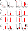
TRP-1 cells expanded in vitro under polarizing conditions show highly different cytokine secretion patterns. (A) Release of INF-γ by Th0, Th1, and Th17 TRP-1 cells as measured by ELISA. Polarized TRP-1–specific TCR transgenic T cells were restimulated overnight with TRP-1106-130 peptide–pulsed splenocytes at escalating concentrations or gp100 control peptide (at a concentration of 10−5 mg/mL; left panel) or were incubated overnight with B16 and B16/CIITA melanoma cell lines. Tumor cell lines lacking the relevant antigen, MCA205 and EL-4, were used as specificity control (right panel). (B) Release of IL-17A by Th0, Th1, and Th17 TRP-1 cells upon stimulation with escalating concentrations of TRP-1106-130 peptide (left) or B16 and B16/CIITA melanoma cell lines (right). MCA205 and EL-4 were used as a negative control. (C) Secretion of TNF-α, IL-2, IL-6, IL-10, IL-17, IL-21, and CCL20 was measured by ELISA after overnight stimulation with TRP-1106-130 peptide–pulsed splenocytes (left panels) or B16 and B16/CIITA melanoma cells (right panels). Splenocytes pulsed with gp-100 peptide and MCA205 and EL-4 tumor cells were used as specificity control.
Figure 3
Figure 3
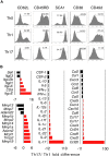
In vitro–polarized effector CD4+ T-cell subsets acquire distinct phenotypes and gene expression profiles. (A) In vitro polarizing conditions alter the phenotype of TRP-1 cells. Th0, Th1, and Th17 TRP-1 T cell were analyzed using flow cytometry for the expression of selected activation markers and adhesion molecules: CD62L, CD45RB, SCA1 (Ly6a), CD38, and CD49d (integrin α4, VLA-4). Percentage of positive cells is calculated based on the comparison with an isotype control antibody. (B) Relative alterations in mRNA quantities of selected genes are observed using microarray analysis. Th1 versus Th17 TRP-1 cells were compared, using Th0 mRNA as a reference. mRNAs are grouped by their function (integrins and adhesion molecules, matrix metalloproteinases [MMPs] and related molecules, cytokines and their receptors, chemokines, and chemokine receptors). A cutoff for significant difference in the level of mRNA message expression was set at 2-fold.
Figure 4
Figure 4
Th17-polarized TRP-1 cells are highly efficient in mediating the rejection of established B16 melanoma tumor upon adoptive cell transfer. (A) C57BL/6 mice B16 tumors that were sublethally irradiated (5 Gy TBI) were left untreated as controls (NT) or received adoptive transfer of 1 × 10⁶ Th0-, Th1-, or Th17-polarized TRP-1 T cells. Th17-treated animals displayed a statistically significant greater tumor regression compared with other groups (P = .001 vs Th0- and Th1-treated groups) that were not different from NT group (P > .05). Results for tumor area are the mean of measurements from at least 5 mice per group (± SEM). Data shown are representative of multiple independent experiments. (B) Percentage of animals alive following treatment described in panel A (n = 5-7, Th1 vs Th17 P < .001). (C) C57BL/6 mice B16 tumors were sublethally irradiated and left untreated as control (NT) or received adoptive transfer of 1 × 10⁶ Th0-, Th1-, or Th17-polarized TRP-1 T cells. In addition, mice received intravenous dose of recombinant TRP-1 vaccinia virus vaccine (rVV) immediately following cell transfer. IL-2 (36 ng/dose) was injected intraperitoneally twice daily for 3 days. Statistically significant tumor regression compared with NT group was observed in all treatment groups. The group treated with Th17 cells had a response significantly better than the Th0-treated group (P < .01), while there was no statistical difference between Th1- and Th17-injected groups (P = .175, n = 5). (D) Percentage of animals alive following treatment described in panel C (combined data from 2 independent experiments (n = 7–14, Th1 vs Th17; P < .001). (E) Animals surviving treatment with Th1 TRP-1 cells, rVV TRP-1 vaccine, and IL-2 developed less vitiligo than mice treated with Th17 TRP-1 cells, vaccination, and IL-2. Vitiligo score: 0 indicates no vitiligo (wild type); 1, depigmentation detected; 2, more than 10% vitiligo; 3, more than 25% vitiligo; 4, more than 50% vitiligo; and 5, more than 75% vitiligo. Evaluation was performed approximately 3 to 4 months after adoptive cell transfer.
Figure 5
Figure 5
Th17-polarized TRP-1 cells have a survival advantage after adoptive transfer into tumor-bearing hosts. (A) Spleens, lymph nodes, and tumors were harvested on day 6 from nontreated animals (NT) or animals treated with Th0, Th1, or Th17 cells and analyzed by flow cytometry for expression of Vβ14 and CD4. The panel is representative of 4 distinct experiments. (B) The total number of Vβ14+CD4+ cells recovered from spleens of treated animals on days 6 and 12 was calculated as described in “Methods” (± SD, n = 3-4). (C) In vivo proliferation of polarized TRP-1 cells. CFSE-labeled Th0, Th1, or Th17 (Thy1.2+) cells were adoptively transferred into 500 R irradiated B6.PL hosts (Thy1.1+). Splenocytes were harvested on days 3 and day 6 and analyzed by flow cytometry. Histograms show CFSE fluorescence after gating on Thy1.2+ population. Th0 and Th1 displayed a greater dilution of the florescent dye compared with Th17 cells. (D) Polarized TRP-1 T cells (Thy1.2+) were transferred into sublethally irradiated tumor-bearing B6.PL (Thy1.1+) hosts. The frequency of Th1.2+CD4+ cells in tumor-draining lymph nodes pooled from at least 3 animals/group was measured by flow cytometry 6 days after adoptive cell transfer.
Figure 6
Figure 6
Th17-polarized TRP-1 CD4+ T cells reject tumor in an IFN-γ–dependent mechanism. (A) In vivo neutralization of cytokines after adoptive transfer of Th17 TRP-1 cells. Sublethally irradiated (500 R) tumor-bearing C57BL/6 mice were treated with 1 × 10⁶ Th17 TRP-1 cells and injected intraperitoneally every other day with 100 μg neutralizing antibodies directed against IFN-γ, IL-17A, IL-23, or isotype control antibody. Results for tumor area are the mean of measurements from at least 5 mice per group (± SEM). Data shown are representative of 3 independent experiments (NT vs Th17, P = .003; NT vs Th17 anti–IFN-γ, P = .558; Th17 isotype vs Th17 anti–IL-17A, P = .117; Th17 isotype vs Th17 anti–IL-23, P = .754). (B) Tumor-bearing C57BL/6, (C) IFN-γ−/−, and (D) IFN-γ receptor–deficient (IFN-γR−/−) mice were treated with 1 × 10⁶ Th1- and Th17-polarized TRP-1 CD4+ T cells. Tumor growth was measured as described previously (± SEM, n = 5-8; NT vs Th17, P < .001 in C57/BL6 hosts, P < .05 in IFN-γ−/− hosts, and P < .05 in IFN-γR−/− hosts).

Comment in

References

    1. Knutson KL, Disis ML. Tumor antigen-specific T helper cells in cancer immunity and immunotherapy. Cancer Immunol Immunother. 2005;54:721–728. - PMC - PubMed
    1. Gerloni M, Zanetti M. CD4 T cells in tumor immunity. Springer Semin Immunopathol. 2005;27:37–48. - PubMed
    1. Jiang YZ, Barrett J. The allogeneic CD4+ T-cell-mediated graft-versus-leukemia effect. Leuk Lymphoma. 1997;28:33–42. - PubMed
    1. Sprangers B, Van Wijmeersch B, Fevery S, Waer M, Billiau AD. Experimental and clinical approaches for optimization of the graft-versus-leukemia effect. Nat Clin Pract Oncol. 2007;4:404–414. - PubMed
    1. Antony PA, Piccirillo CA, Akpinarli A, et al. CD8+ T cell immunity against a tumor/self-antigen is augmented by CD4+ T helper cells and hindered by naturally occurring T regulatory cells. J Immunol. 2005;174:2591–2601. - PMC - PubMed