Skip to main page content
U.S. flag

An official website of the United States government

Dot gov

The . gov means it’s official VSports app下载. Federal government websites often end in . gov or . mil. Before sharing sensitive information, make sure you’re on a federal government site. .

Https

The site is secure. The https:// ensures that you are connecting to the official website and that any information you provide is encrypted and transmitted securely V体育官网. .

. 2008 Feb;10(2):131-9.
doi: 10.1593/neo.07815.

Endothelial cells enhance tumor cell invasion through a crosstalk mediated by CXC chemokine signaling

Affiliations

"VSports app下载" Endothelial cells enhance tumor cell invasion through a crosstalk mediated by CXC chemokine signaling

"VSports最新版本" Kristy A Warner et al. Neoplasia. 2008 Feb.

Abstract

Field cancerization involves the lateral spread of premalignant or malignant disease and contributes to the recurrence of head and neck tumors. The overall hypothesis underlying this work is that endothelial cells actively participate in tumor cell invasion by secreting chemokines and creating a chemotactic gradient for tumor cells. Here we demonstrate that conditioned medium from head and neck tumor cells enhance Bcl-2 expression in neovascular endothelial cells. Oral squamous cell carcinoma-3 (OSCC3) and Kaposi's sarcoma (SLK) show enhanced invasiveness when cocultured with pools of human dermal microvascular endothelial cells stably expressing Bcl-2 (HDMEC-Bcl-2), compared to cocultures with empty vector controls (HDMEC-LXSN). Xenografted OSCC3 tumors vascularized with HDMEC-Bcl-2 presented higher local invasion than OSCC3 tumors vascularized with control HDMEC-LXSN. CXCL1 and CXCL8 were upregulated in primary endothelial cells exposed to vascular endothelial growth factor (VEGF), as well as in HDMEC-Bcl-2. Notably, blockade of CXCR2 signaling, but not CXCR1, inhibited OSCC3 and SLK invasion toward endothelial cells. These data demonstrate that CXC chemokines secreted by endothelial cells induce tumor cell invasion and suggest that the process of lateral spread of tumor cells observed in field cancerization is guided by chemotactic signals that originated from endothelial cells VSports手机版. .


VSports app下载 - Figures

Figure 1
Figure 1
Upregulated Bcl-2 expression in endothelial cells enhances tumor cell invasion in vitro. (A) Western blot analysis of Bcl-2 expression in HDMECs exposed to 0 or 50 ng/ml rhVEGF165 for 24 hours, or in HDMEC exposed to tumor cell.conditioned medium from UM-SCC-17B, OSCC3, SLK, and UM-SCC-74A cells for 24 hours using a monoclonal anti-Bcl-2 antibody. (B) Western blot analysis to characterize Bcl-2 expression in three independent pools of HDMEC stably transduced with Bcl-2, or with empty retroviral vector (LXSN) using a mouse anti-Flag antibody. (C) Graph depicting OSCC3 or SLK invasion toward HDMEC-LXSN or HDMEC-Bcl-2 cells in vitro. *P ≤ .05. Data represent mean values (± SD) of three independent experiments.
Figure 2
Figure 2
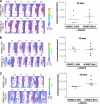
Evaluation of tumor progression over time by in vivo bioluminescence. (A) Experimental design used for acquisition of data presented in Figures 2 and 3. (B) In vivo bioluminescent images of one mouse implanted with HDMEC-LXSN + OSCC3 cells and one mouse implanted with HDMEC-Bcl-2 + OSCC3 cells over time (i.e., 14, 28, 34, 41 and 56 days after implantation). (C) Graph depicting in vivo bioluminescent values after 56 days. *P ≤ .05. Data presented in C correspond to the analysis of six mice per condition.
Figure 3
Figure 3
Upregulation of Bcl-2 expression in endothelial cells enhances xenograft tumor recurrence. (A–C) In vivo bioluminescent images of each mouse implanted with HDMEC-LXSN + OSCC3 cells and each mouse implanted with HDMEC-Bcl-2 + OSCC3 cells; and corresponding scatter plots showing each individual data point and the median bioluminescence value (horizontal line). (A) Bioluminescence of primary tumors (28 days). (B) Bioluminescence immediately after removal of the primary tumors (35 days). (C) Bioluminescence of the local recurrences (56 days).
Figure 4
Figure 4
Primary and recurrent xenograft tumors show similar histologic features. Hematoxylin and eosin staining of a (A) primary and (B) recurrent tumor retrieved from a mouse implanted with HDMEC-LXSN + OSCC3 cells. (C) Primary and (D) recurrent tumor retrieved from a mouse implanted with HDMEC-Bcl-2 + OSCC3 cells. Original magnification, 400x.
Figure 5
Figure 5
Head and neck tumor cells express CXCR1 and CXCR2. (A) Graph showing CXCL8 and (B) CXCL1 expression in endothelial cells treated with 50.0 ng/ml rhVEGF165 for 24 hours, two independent pools of HDMEC-Bcl-2, HDMEC-LXSN, or untreated controls. *P ≤ .05 relative to untreated controls. (C) Western blot analysis of CXCR1 and CXCR2 expression in endothelial and a panel of tumor cells.
Figure 6
Figure 6
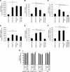
Blockade of CXCR2, but not CXCR1, signaling prevents tumor cell invasion toward endothelial cells expressing Bcl-2. (A–F) Graphs characterizing the effect of blockade of CXCR1 and/or CXCR2 signaling on (A–C) SLK, or (D–F) OSCC3 tumor cell invasion in vitro. Cells were exposed to 1.0 µg/ml anti-CXCR1 (A and D), 1.0 µg/ml anti-CXCR2 (B and E), or to both anti-CXCR1 and anti-CXCR2 (C and F) at the same time. As controls, we exposed cells to 1.0 µg/ml nonspecific isotype-matched IgG, or left cells untreated. Data represent mean values (± SD) of three independent experiments. Statistical significance (P ≤ .05) is depicted by lowercase letters: a tumor cell invasion is significantly higher toward HDMEC-Bcl-2 or HDMEC-Bcl-2 + IgG than toward HDMEC-LXSN; b tumor cell invasion is significantly higher toward HDMEC-Bcl-2 or HDMEC-Bcl-2 + IgG than toward HDMEC-Bcl-2 + anti-CXCR2 or HDMEC-Bcl-2 + anti-CXCR1 + anti-CXCR2. (G) SRB analysis of cells exposed to the same conditions as above for determination of the effects of the antibodies on cell number. Data represent mean values (± SD) of two independent experiments.

References

    1. Davies L, Welch HG. Epidemiology of head and neck cancer in the United States. Otolaryngol Head Neck Surg. 2006;135:451–457. - "VSports最新版本" PubMed
    1. de Vicente JC, Olay S, Lequerica-Fernandez P, Sanchez-Mayoral J, Junquera LM, Fresno MF. Expression of Bcl-2 but not Bax has a prognostic significance in tongue carcinoma. J Oral Pathol Med. 2006;35:140–145. - "V体育平台登录" PubMed
    1. Genden EM, Ferlito A, Bradley PJ, Rinaldo A, Scully C. Neck disease and distant metastases. Oral Oncol. 2003;39:207–212. - "V体育官网入口" PubMed
    1. O'Donnell RK, Kupferman M, We SJ, Singhal S, Weber R, O'Malley B, Cheng Y, Putt M, Feldman M, Ziober B, et al. Gene expression signature predicts lymphatic metastasis in squamous cell carcinoma of the oral cavity. Oncogene. 2005;24:1244–1251. - PubMed
    1. Slaughter DP, Southwick HW, Smejkal W. “Field cancerization” in oral stratified squamous epithelium. Cancer. 1953;6:963–968. - PubMed

Publication types

Substances (VSports注册入口)