Skip to main page content
U.S. flag

An official website of the United States government

Dot gov

The . gov means it’s official. Federal government websites often end in . gov or . mil VSports app下载. Before sharing sensitive information, make sure you’re on a federal government site. .

Https

The site is secure V体育官网. The https:// ensures that you are connecting to the official website and that any information you provide is encrypted and transmitted securely. .

. 2008 Apr 15;177(8):861-70.
doi: 10.1164/rccm.200708-1269OC. Epub 2008 Jan 3.

SIRT1, an antiinflammatory and antiaging protein, is decreased in lungs of patients with chronic obstructive pulmonary disease

Affiliations

SIRT1, an antiinflammatory and antiaging protein, is decreased in lungs of patients with chronic obstructive pulmonary disease (VSports app下载)

Saravanan Rajendrasozhan et al. Am J Respir Crit Care Med. .

Abstract

Rationale: Abnormal inflammation and accelerated decline in lung function occur in patients with chronic obstructive pulmonary disease (COPD). Human sirtuin (SIRT1), an antiaging and antiinflammatory protein, is a metabolic NAD(+)-dependent protein/histone deacetylase that regulates proinflammatory mediators by deacetylating histone and nonhistone proteins. VSports手机版.

Objectives: To determine the expression of SIRT1 in lungs of smokers and patients with COPD, and to elucidate the regulation of SIRT1 in response to cigarette smoke in macrophages, and its impact on nuclear factor (NF)-kappaB regulation V体育安卓版. .

Methods: SIRT1 and NF-kappaB levels were assessed in lung samples of nonsmokers, smokers, and patients with COPD. Human monocyte-macrophage cells (MonoMac6) were treated with cigarette smoke extract (CSE) to determine the mechanism of CSE-mediated regulation of SIRT1 and its involvement in RelA/p65 regulation and IL-8 release V体育ios版. .

Measurements and main results: Peripheral lungs of smokers and patients with COPD showed decreased levels of nuclear SIRT1, as compared with nonsmokers, associated with its post-translational modifications (formation of nitrotyrosine and aldehyde carbonyl adducts) VSports最新版本. Treatment of MonoMac6 cells with CSE showed decreased levels of SIRT1 associated with increased acetylation of RelA/p65 NF-kappaB. Mutation or knockdown of SIRT1 resulted in increased acetylation of nuclear RelA/p65 and IL-8 release, whereas overexpression of SIRT1 decreased IL-8 release in response to CSE treatment in MonoMac6 cells. .

Conclusions: SIRT1 levels were reduced in macrophages and lungs of smokers and patients with COPD due to its post-translational modifications by cigarette smoke-derived reactive components, leading to increased acetylation of RelA/p65 V体育平台登录. Thus, SIRT1 plays a pivotal role in regulation of NF-kappaB-dependent proinflammatory mediators in lungs of smokers and patients with COPD. .


Figures

<b>Figure 1.</b>
Figure 1.
Decreased levels of sirtuin (SIRT1) protein in lung tissue of smokers and patients with chronic obstructive pulmonary disease (COPD). (A) Western blot analysis of SIRT1 in soluble nuclear proteins (30 μg) extracted from the lung tissue of nonsmokers (n = 10), smokers (n = 10), and patients with COPD (n = 17). The proteins were electrophoresed on a 7.5% polyacrylamide gel electrophoresis and electroblotted onto a nitrocellulose membrane. The level of SIRT1 protein was determined using mouse monoclonal anti-SIRT1 antibody. The purity of nuclear extract was shown by the presence of lamin B (nuclear envelope protein) and the absence of the cytoskeletal protein α-tubulin (not shown). (B) After densitometric analysis, the values were normalized against the loading control, β-actin. The relative level (% of control) of SIRT1 showed decreased levels of nuclear SIRT1 protein in the lung tissues of smokers and patients with COPD. (C) SIRT1 protein was immunoprecipitated from the nuclear extract of lung homogenates. The levels of SIRT1 adducts with 4-hydroxy-2-nonenol (4-HNE) and nitration of tyrosine residues on SIRT1 were analyzed by immunoblotting with anti–4-HNE and anti–3-nitrotyrosine (3-NT) antibodies, respectively. Equal amount of immunoprecipitated SIRT1 protein (100 μg) was used for Western blotting. (D) Relative intensity of 4-HNE/SIRT1 and 3-NT/SIRT1 represents the increased post-translational modifications of SIRT1 protein in lungs of smokers and patients with COPD compared with nonsmokers. A representative blot is shown, which was obtained from several blotting experiments. Results are expressed as mean ± SEM. ***P < 0.001, significant compared with nonsmokers.
<b>Figure 2.</b>
Figure 2.
Decreased staining of sirtuin (SIRT1) in lung macrophages and alveolar/airway epithelial cells of smokers with and without chronic obstructive pulmonary disease (COPD). (A) Representative photographs (original magnification, ×400) from immunostaining for SIRT1 in lung tissues from nonsmokers (n = 10) and smokers with (n = 17) and without (n = 10) COPD. The levels of SIRT1 were measured in the fixed lung sections (3-μm thick) by immunohistochemical staining using rabbit polyclonal anti-SIRT1 antibody (1:100 dilution) with avidin–biotin–peroxidase complex method followed by hematoxylin counter staining. Dark brown color represents the presence of SIRT1 (indicated with thick arrow), which was decreased in smokers' lung (indicated with thin arrow). Alv = alveoli; Aw = airway; E = epithelial cells; M = macrophage. (B) Immunostaining scores for SIRT1 per cell type in alveolar and airway regions of the lung. The assessment of immunostaining intensity was performed semiquantitatively and in a blinded fashion. Solid bars, intense staining; shaded bars, moderate/weak staining; open bars, no staining. Results are represented as mean ± SEM. ***P < 0.001, significant compared with nonsmokers.
<b>Figure 3.</b>
Figure 3.
Decreased levels of sirtuin (SIRT1) in smokers and patients with chronic obstructive pulmonary disease (COPD) were associated with increased levels of RelA/p65 nuclear factor (NF)-κB. The expression of NF-κB was measured in fixed lung sections (3-μm thick) of nonsmokers (n = 10), and smokers with (n = 17) and without (n = 10) COPD by immunohistochemical staining using anti-RelA/p65 NF-κB antibody (1:100 dilution) followed by incubations with fluorescein isothiocyanate conjugated anti-rabbit secondary antibody. Representative immunofluorescent images (original magnification, ×400) showed increased levels of NF-κB in the lungs (especially in macrophages and epithelial cells) of smokers with and without COPD as compared with nonsmokers. Arrows indicate the cells (airway/alveolar epithelial cells and macrophages) that express increased levels of RelA/p65 NF-κB proteins. (B) Immunostaining score for RelA/p65 was performed semiquantitatively and in a blinded fashion: 0 = no staining, 1 = weak staining, 2 = moderate staining, 3 = intense staining. Results are expressed as mean ± SEM. ***P < 0.001, significant compared with nonsmokers.
<b>Figure 4.</b>
Figure 4.
Decreased levels of sirtuin (SIRT1) protein and mRNA expression by cigarette smoke extract (CSE) treatment in monocyte–macrophage (MonoMac6) cells. (A) Western blots of soluble nuclear proteins (30 μg) extracted from CSE-exposed MonoMac6 cells. Expression of SIRT1 was determined using mouse monoclonal anti-SIRT1 antibody. The purity of nuclear extract was shown by the presence of lamin B (nuclear envelope protein) and the absence of the cytoskeletal protein α-tubulin (bands not shown). Gel pictures shown are representative of at least three separate experiments. (B) Densitometric values of SIRT1 were normalized against the loading control, β-actin. The relative level (% of control) of SIRT1 in MonoMac6 cells showed decreased levels of SIRT1 protein in response to CSE at 4 and 24 hours. (C) CSE decreased the levels of SIRT1 mRNA in MonoMac6 cells. After 4 and 24 hours of CSE treatment, total RNA was extracted from MonoMac6 cells using RNeasy kit (Qiagen, Germantown, MD). Reverse transcriptase–polymerase chain reaction was performed. Amplified products (SIRT1: 200 bp; GAPDH: 600 bp) were resolved by 1.5% agarose gel electrophoresis, stained with ethidium bromide. SIRT1 mRNA expression was decreased after 24-hour exposure to low concentrations of CSE (0.5 and 1%) compared with control. Data represent mean ± SEM of three experiments (n = 3). *P < 0.05, **P < 0.01, ***P < 0.001, significant compared with control values.
<b>Figure 5.</b>
Figure 5.
Decreased sirtuin (SIRT1) protein staining in response to cigarette smoke extract (CSE) treatment in monocyte–macrophage (MonoMac6) cells. (A) CSE decreased the levels of SIRT1 in MonoMac6 cells at 4 and 24 hours. MonoMac6 cells were treated with different concentrations of CSE (0.1–1.0%). Cells were harvested and the cytospin slides were prepared at 4 and 24 hours of treatments. Immunostaining was performed using a rabbit polyclonal antibody specific for SIRT1 followed by the avidin–biotin–peroxidase complex method, and counterstained with hematoxylin. The dark brown color represents the presence of SIRT1, which was decreased in response to CSE treatment. (B) Graph showing the percentage of SIRT1-positive cells from the total number of cells in CSE-treated MonoMac6 cells. The assessment of immunostaining intensity was performed semiquantitatively and in a blinded fashion. Results shown are means ± SEM of three separate experiments (n = 3). ***P < 0.001, significant compared with control values.
<b>Figure 6.</b>
Figure 6.
Cigarette smoke extract (CSE) induced IL-8 release from monocyte–macrophage (MonoMac6) cells. MonoMac6 cells were treated with freshly prepared CSE (0.1, 0.5, and 1.0%) for 4 and 24 hours. IL-8 release was measured in the culture media by sandwich ELISA duo-antibody kit (R&D Systems, Minneapolis, MN). CSE showed increase in the levels of IL-8 as compared with controls at 4 and 24 hours. Each histogram represents the mean ± SEM of three experiments (n = 3). **P < 0.01, ***P < 0.001, significant compared with controls.
<b>Figure 7.</b>
Figure 7.
Cigarette smoke extract (CSE)–mediated IL-8 release was modified by sirtuin (SIRT1) knockdown, mutation, and overexpression in monocyte–macrophage (MonoMac6) cells. (A) MonoMac6 cells were transfected with predesigned human SIRT1 siRNA or scrambled nontarget siRNA using DharmaFect2 siRNA transfection reagent. After 36–48 hours of transfection (cells > 85% viable), the cells were treated with CSE (0.5%) for 12 hours. At the end of the experiment, culture media was collected by centrifugation for IL-8 assay. SIRT1 knockdown led to significant increase in IL-8 release in response to CSE treatment in MonoMac6 cells as compared with nontarget scrambled siRNA. (B) MonoMac6 cells were transfected with SIRT1 overexpressing plasmid or SIRT1-H363Y (mutated in the deacetylase domain) using the calcium phosphate method. Overexpression of SIRT1 deceased IL-8 release, whereas SIRT1-H363Y lacking SIRT1 deacetylase domain increased IL-8 release in response to CSE treatment at 4 hours. Each value is the mean ± SEM of triplicate determinations (n = 3). **P < 0.001, ***P < 0.001, significant compared with control; ##P < 0.01, ###P < 0.001, significant compared with CSE-treated group.
<b>Figure 8.</b>
Figure 8.
Cigarette smoke extract (CSE) caused post-translational modifications of sirtuin (SIRT1). (A) SIRT1 protein was immunoprecipitated from the nuclear extract of monocyte–macrophage (MonoMac6) cells treated with CSE (0.1, 0.5, and 1.0%) for 4 hours. The levels of SIRT1 adducts with 4-hydroxy-2-nonenol (4-HNE) and nitration of tyrosine residues on SIRT1 were analyzed by immunoblotting with anti–4-HNE and anti–3-nitrotyrosine (3-NT) antibodies, respectively. Equal amount of immunoprecipitated SIRT1 protein (100 μg) was used for Western blotting. Relative intensity of 4-HNE/SIRT1 (B) and 3-NT/SIRT1 (C) represents the increased post-translational modifications of SIRT1 protein in response to CSE treatment. Results are means ± SEM of three separate experiments (n = 3). Significant differences are shown as compared to controls: *P < 0.05, **P < 0.01, and ***P < 0.001.
<b>Figure 9.</b>
Figure 9.
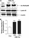
Cigarette smoke extract (CSE)–mediated decrease in sirtuin (SIRT1) level was associated with increased acetylation of RelA/p65 nuclear factor (NF)-κB. (A) Monocyte–macrophage (MonoMac6) cells were treated with CSE (0.5 and 1.0%) for 4 hours. Acetylation of the lysine residue (K310) on RelA/p65 NF-κB protein was determined in soluble nuclear proteins (30 μg) by Western blotting using anti–acetyl RelA/p65 (K310) antibody. β-Actin was measured as a loading control. Lamin B (nuclear envelope protein) and the absence of the cytoskeletal protein α-tubulin (bands not shown) were measured to confirm the purity of nuclear extracts. (B) The relative density (% of control) of acetylated RelA/p65 NF-κB in nuclear fraction of MonoMac6 cells showed increased acetylation of RelA/p65 NF-κB in response to CSE treatment at 4 hours. Each histogram represents the means ± SEM (n = 3). ***P < 0.001, compared with control values.
<b>Figure 10.</b>
Figure 10.
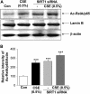
siRNA silencing of sirtuin (SIRT1) augmented the cigarette smoke extract (CSE)–mediated acetylation of RelA/p65 nuclear factor (NF)-κB. Monocyte–macrophage (MonoMac6) cells were transfected with predesigned human SIRT1 siRNA duplex (100 nM) using DharmaFect2 transfection reagent for 36–48 hours and then treated with CSE (0.5%) for 12 hours. Nontargeting scrambled siRNA was used as a negative control. Actin was measured as a loading control. (A) Acetylation of RelA/p65 NF-κB was determined using rabbit anti-Ac-RelA/p65 (K310) antibody in the soluble nuclear extract. The purity of nuclear extract was shown by the presence of lamin B (nuclear envelope protein) and the absence of the cytoskeletal protein α-tubulin (bands not shown). (B) The relative level (% of control) of Ac-RelA/p65 showed increased acetylation of nuclear RelA/p65 in response to SIRT1 knockdown and/or CSE treatment. Each value is the mean ± SEM of triplicate determinations (n = 3). ***P < 0.001, significant compared with control.

References (VSports)

    1. Pauwels RA, Buist AS, Calverley PM, Jenkins CR, Hurd SS. Global strategy for the diagnosis, management, and prevention of chronic obstructive pulmonary disease. NHLBI/WHO Global Initiative for Chronic Obstructive Lung Disease (GOLD) workshop summary. Am J Respir Crit Care Med 2001;163:1256–1276. - V体育ios版 - PubMed
    1. Murray CJ, Lopez AD. Mortality by cause for eight regions of the world: Global Burden of Disease Study. Lancet 1997;349:1269–1276. - PubMed
    1. Vogelmeier C, Bals R. Chronic obstructive pulmonary disease and premature aging. Am J Respir Crit Care Med 2007;175:1217–1218. - V体育官网入口 - PubMed
    1. Tuder RM. Aging and cigarette smoke: fueling the fire. Am J Respir Crit Care Med 2006;174:490–491. - "V体育平台登录" PubMed
    1. Barnes PJ. Chronic obstructive pulmonary disease. N Engl J Med 2000;343:269–280. - PubMed

Publication types

"V体育2025版" MeSH terms

V体育官网入口 - Grants and funding