Skip to main page content
U.S. flag

An official website of the United States government

Dot gov

The . gov means it’s official. Federal government websites often end in . gov or VSports app下载. mil. Before sharing sensitive information, make sure you’re on a federal government site. .

Https

The site is secure. The https:// ensures that you are connecting to the official website and that any information you provide is encrypted and transmitted securely. V体育官网.

. 2007 Dec 24;204(13):3221-34.
doi: 10.1084/jem.20071285. Epub 2007 Dec 3.

Antiribosomal-P autoantibodies from psychiatric lupus target a novel neuronal surface protein causing calcium influx and apoptosis (VSports手机版)

Affiliations

Antiribosomal-P autoantibodies from psychiatric lupus target a novel neuronal surface protein causing calcium influx and apoptosis

Soledad Matus et al. J Exp Med. .

Abstract

The interesting observation was made 20 years ago that psychotic manifestations in patients with systemic lupus erythematosus are associated with the production of antiribosomal-P protein (anti-P) autoantibodies. Since then, the pathogenic role of anti-P antibodies has attracted considerable attention, giving rise to long-term controversies as evidence has either contradicted or confirmed their clinical association with lupus psychosis. Furthermore, a plausible mechanism supporting an anti-P-mediated neuronal dysfunction is still lacking. We show that anti-P antibodies recognize a new integral membrane protein of the neuronal cell surface VSports手机版. In the brain, this neuronal surface P antigen (NSPA) is preferentially distributed in areas involved in memory, cognition, and emotion. When added to brain cellular cultures, anti-P antibodies caused a rapid and sustained increase in calcium influx in neurons, resulting in apoptotic cell death. In contrast, astrocytes, which do not express NSPA, were not affected. Injection of anti-P antibodies into the brain of living rats also triggered neuronal death by apoptosis. These results demonstrate a neuropathogenic potential of anti-P antibodies and contribute a mechanistic basis for psychiatric lupus. They also provide a molecular target for future exploration of this and other psychiatric diseases. .


Figures

Figure 1.
Figure 1.
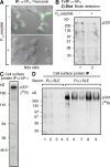
Affinity-purified anti-P antibodies recognize a cell-surface protein of high molecular mass (p331) in neurons. (A) Immunoblot (IB) with affinity-purified anti-P antibodies (α-hP11) shows reaction with the P ribosomal proteins P0 (38 kD), P1 (19 kD), and P2 (17 kD; lane 1), which is blocked by the P peptide (lane 2). (B) Anti-P staining of the neuronal surface. Brain cortical primary culture cells were incubated with α-hP11 antibody at 4°C, and fixed and incubated with secondary FITC–anti–human antibody. Some neurons show intense surface staining. Bar, 10 μm. (C) Detection of anti-P target at the surface of brain cortical neurons. Neurons in primary culture were metabolically labeled for 16 h with 100 μCi [35S]methionine-cysteine, biotinylated at 4°C, and subjected to immunoprecipitation with either control P (−) serum from an SLE patient (lane 1) or α-hP11 antibodies (lane 2). The immunoprecipitated proteins were separated from the sepharose beads by heating in SDS buffer, and the biotinylated proteins were precipitated with immobilized neutravidin protein. A single high molecular mass protein (p331) is detected.
Figure 2.
Figure 2.
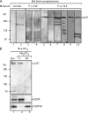
Antibodies against ribosomal P epitope recognize p331 in the surface of N2a cells. (A) Anti-P indirect immunofluorescence in intact neuroblastoma N2a cells. The cell-surface staining, superimposed over images of Nomarski interference microscopy, is asymmetrically distributed and is displaced by the P peptide. Bar, 10 μm. (B) Biotinylation assays detect p331 as the cell-surface P epitope–bearing protein. Intact N2a cells were biotinylated at 4°C, lysed, and subjected to immunoprecipitation with α-hP11 antibodies either in the absence (lane 1) or presence (lane 2) of P peptide. The biotinylated proteins, resolved by SDS-PAGE, were detected in the blot with horseradish peroxidase–streptavidin. (C and D) Direct immunoprecipitation of cell-surface p331 anti-P target. N2a cells metabolically labeled for 16 h with 100 μCi [35S]methionine-cysteine were incubated intact, at 4°C, with either α-hP11 (C) or sera from lupus patients (D), including anti-P (−; lanes 1–4) or anti-P (+) with psychosis (lane 5) or without NP-SLE (lanes 6–9). The cells were lysed, and the immunocomplexes formed at the cell surface were precipitated with protein A–sepharose, resolved by SDS-PAGE, and visualized by fluorography, showing only the p331 protein.
Figure 3.
Figure 3.
The anti-P target p331 is an integral membrane protein present in rat brain synaptosomes. (A) Detection of p331 in rat brain synaptosomes. Membrane fractions prepared from rat brain synaptosomes were analyzed by immunoblot (IB) with control sera from healthy individuals (lanes 1 and 2), anti-P (−) from two SLE patients (lanes 3 and 4), and anti-P (+) from six SLE patients (lanes 5–10), including one with psychosis (lane 10). All P (+) serum detected p331, providing further evidence of its expression in brain neurons. (B) p331 is an integral membrane protein and the main α-hP11 target in synaptosomes. Immunoblot of synaptosomes (Syn) with affinity-purified α-hP11 antibodies shows p331 as the main target (lane 1). The lower bands are degradation products that vary in different experiments. Sodium carbonate extraction of synaptosomal membranes shows that p331 distributes exclusively to the pellet (P; lane 2), similar to the integral membrane protein dopamine receptor (D2DR), whereas peripherally associated clathrin distributes to the supernatant (SN; lane 3).
Figure 4.
Figure 4.
p331 is a novel protein named NSPA. (A) Preparative immunoprecipitation of neuronal anti-P target. Presynaptosomal fractions (P2) obtained from 50 rat brains were subjected to immunoprecipitation with affinity-purified anti-P antibodies. SDS-PAGE and Coomassie blue staining show two main bands (asterisks; IgG-HC, IgG–heavy chain) that were analyzed by mass spectrometry and microsequencing. The 180-kD protein corresponds to clathrin, whereas p331 corresponds to the human protein NP055928 of unknown function. (B) Sequence of p331-derived peptides, possible P epitopes, and the immunogenic NSPA peptide. Six out of six peptides derived from p331 (P1–6) matched perfectly with the putative human protein NP055928 at the indicated regions. We named this protein NSPA. The NSPA sequences 644DDLG647 and 2884GLFD2887 are homologous to the ribosomal DDMG and GLFD P epitopes within the 11 C-terminal region. The peptide sequence 2876–2886 used to immunize rabbits to obtain anti-NSPA antibodies is also shown.
Figure 5.
Figure 5.
Two distant regions of NSPA containing 644DDLG647 and 2881GLFD2884 sequences participate in the cell-surface interaction with anti-P autoantibodies. (A) Blocking activity of synthetic peptides against the cell-surface staining of α-hP11 antibodies involved both 644DDLG647 and 2881GLFD2884 NSPA sequences. Indirect immunofluorescence with α-hP11 in nonpermeabilized N2a cells either in the absence or presence of NSPA peptides PDDLG (642SSDDLGED649) and PGLFE (2876THMEYGLFEDV2886) or their corresponding scrambled peptides, randomized PDDLG (LGDSSEDLD) and randomized PGLFE (DEYTEHFGLVM). Both PDDLG and PGLFE peptides, but not their randomized versions, decreased the cell-surface α-hP11 immunostaining. An IgG fraction isolated from a P (−) serum gives no staining. In all of the images, some staining background is depicted on purpose to visualize the cells. Bar, 10 μm. (B–D) A subset of anti-P antibodies (α-hP4) recognize P ribosomal proteins and NSPA. A P (+) serum was subjected to affinity chromatography with the NSPA peptide DDLGEDD containing a putative P epitope (DDLG). The isolated α-hP4 antibodies, similar to the α-hP11 antibodies, recognize in immunoblot (IB) the P ribosomal proteins in ribosomes isolated from rat liver (B), and the high molecular mass protein (p331) the in synaptosomes (Syn; lane 1) and pellet (P; lane 2) but not the supernatant (SN; lane 3) of carbonate-extracted membranes (C). (D) Indirect immunofluorescence with α-hP4 antibodies shows the characteristic asymmetrical cell-surface staining, which was abrogated by the PDDLG (642SSDDLGED649) peptide. Bar, 10 μm.
Figure 6.
Figure 6.
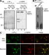
Polyclonal antibodies against NSPA. (A) Immunoblots (IB) with human α-hP11 autoantibodies and rabbit anti-NSPA (α-NSPA) antibodies of carbonate-extracted synaptosomal membranes. Both antibodies recognize the high molecular mass protein (p331/NSPA) in total the synaptosomal membranes (Syn; lanes 1 and 4) and pellet (P; lanes 2 and 5) but not the supernatant (SN; lanes 3 and 6) of carbonate extraction. (B) Immune cross reaction of NSPA with α-hP11 and α-NSPA antibodies. The p331/NSPA protein immunoprecipitated (IP) by α-hP11 from extracts of a synaptosomes (lane 1) is recognized in immunoblot by α-NSPA but not by the preimmune serum (PI; lane 2). (C) Double immunofluorescence with α-NSPA and α-hP11 antibodies. Nonpermeabilized N2a cells and cortical neurons in primary culture both show complete cell-surface colocalization of α-NSPA (red) and α-hP11 (green) staining. All of these results indicate that α-NSPA and α-hP11 antibodies recognize the same protein. Bar, 10 μm.
Figure 7.
Figure 7.
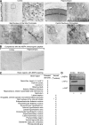
NSPA is expressed by neurons at specific regions of the brain. (A) Immunohistochemistry of NSPA in the rat brain. Some brain regions with framed zones of higher magnification (insets) are illustrated. Arrows indicate interneurons, synaptic buttons, and calyx-like terminals. Bars: (cortex, left) 100 μm; (cortex, right) 10 μm; (hippocampus, left) 50 μm; (hippocampus, right) 10 μm. The bed nucleus of stria terminalis and the central nucleus of the amygdala are also shown. Bars, 10 μm. (B) The immunogenic NSPA peptide completely abrogated the staining (not depicted). (C) Summary of NSPA expression in neurons of different areas of the brain, indicating whether the staining was detected in somatodendritic or axonal (synaptic buttons) regions. (D) Correspondence between immunohistochemical staining and immunoblot detection of NSPA. Synaptosomes were isolated from the cortex (lane 1), which shows positive immunohistochemical staining, and from the striatum (lane 2), which completely lacks staining. Immunoblot with α-NSPA and α-hP11 show p331 only in synaptosomes from the cortex. In contrast, the dopamine transporter (DAT), which is 90% expressed in the striatum (reference 73), was exclusively detected in this region, thus showing that these two brain areas were effectively isolated.
Figure 8.
Figure 8.
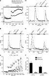
Anti-P and anti-NSPA antibodies induce calcium influx in cortical primary neurons. Graphs illustrate cells displaying different degrees of responsiveness. (A) α-hP11 antibodies induce a sustained increase in calcium levels. The cells were incubated with 0.1 μg/ml (gray bar) and 0.2 μg/ml (black bar) of α-hP11 antibodies. (B) Neuronal responses to α-hP11 are specific and reversible. The increased calcium levels elicited by α-hP11 returned to basal levels after washing the antibody (white bar) and were abrogated by preincubating the antibodies with the P peptide (black bar). (C) Dose-dependent responses to α-hP11. The same cell showed increased responses to increasing amounts of antibody added after a wash out. Horizontal bars represent incubation time periods. (D) Chelation of extracellular Ca2 + with 10 mM EGTA abolished the effects of α-hP11. (E) 1 μg/ml of α-NSPA antibodies also provoke an increase in neuronal calcium levels of lower magnitude, which is reversible, specifically inhibited by the corresponding immunogenic peptide, and abolished by EGTA. (F) Neurons predominantly responded to both α-hP11 and α-NSPA. Although 67 and 51% of neurons increased their calcium levels in response to either α-hP11 or α-NSPA, respectively, only 5 and 3% of the astrocytes showed detectable responses. Control experiments showed that astrocytes responded to glutamate (not depicted). Error bars represent the mean ± SEM.
Figure 9.
Figure 9.
α-hP11 autoantibodies induce neuronal apoptosis in vitro and in vivo. (A) α-hP11 antibodies induce apoptosis in neurons in primary culture. Cells were incubated with 100 μg/ml of control IgG or 0.5 μg/ml of α-hP11 for 48 h. Indirect immunofluorescence against activated caspase-3 revealed apoptosis. (B) α-hP11 antibodies decrease neuronal viability. MTT assays show a concentration-dependent loss of viability. In contrast with the 30% viability loss caused by 0.5 μg/ml of an IgG fraction from a normal individual, the same concentration of α-hP11 lowered the viability by ∼90%. For comparison, H2O2 at 50 μM decreased the viability by 80%. Error bars represent the mean ± SEM. (C) α-hP11 antibodies induce apoptosis in neurons in situ. The rat brain cortex was injected with 0.7 μg of either control IgG or α-hP11 in contralateral sides, fixed after 24 h, and either stained with Cresyl violet or treated for immunohistochemistry of activated caspase-3. The region injected with α-hP11 shows caspase-3 activation (bottom right and inset) and lower cell density (top right) and. Bars, 100 μm.

References

    1. Sherer, Y., A. Gorstein, M.J. Fritzler, and Y. Shoenfeld. 2004. Autoantibody explosion in systemic lupus erythematosus: more than 100 different antibodies found in SLE patients. Semin. Arthritis Rheum. 34:501–537. - PubMed
    1. West, S.G. 2007. The nervous system. In Dubois' Lupus Erythematosus. Seventh edition. D.J. Wallace and B.H. Hahn, editors. Lippincott Williams & Wilkins, Philadelphia. 707–746.
    1. Hanly, J.G., A. Kuznetsova, and J.D. Fisk. 2007. Psychopathology of lupus and neuroimaging. In Dubois' Lupus Erythematosus. Seventh edition. D.J. Wallace and B.H. Hahn, editors. Lippincott Williams & Wilkins, Philadelphia. 747–774.
    1. Hanly, J.G., M.B. Urowitz, J. Sanchez-Guerrero, S.C. Bae, C. Gordon, D.J. Wallace, D. Isenberg, G.S. Alarcon, A. Clarke, S. Bernatsky, et al. 2007. Neuropsychiatric events at the time of diagnosis of systemic lupus erythematosus: an international inception cohort study. Arthritis Rheum. 56:265–273. - PubMed
    1. Karassa, F.B., A. Afeltra, A. Ambrozic, D.M. Chang, F. De Keyser, A. Doria, M. Galeazzi, S. Hirohata, I.E. Hoffman, M. Inanc, et al. 2006. Accuracy of anti-ribosomal P protein antibody testing for the diagnosis of neuropsychiatric systemic lupus erythematosus: an international meta-analysis. Arthritis Rheum. 54:312–324. - PubMed

Publication types (VSports注册入口)

V体育平台登录 - MeSH terms