Skip to main page content
U.S. flag

An official website of the United States government

Dot gov

The . gov means it’s official. Federal government websites often end in . gov or VSports app下载. mil. Before sharing sensitive information, make sure you’re on a federal government site. .

Https

The site is secure. The https:// ensures that you are connecting to the official website and that any information you provide is encrypted and transmitted securely V体育官网. .

. 2007 Dec;22(12):1957-67.
doi: 10.1359/jbmr.070804.

"VSports在线直播" Control of the SOST bone enhancer by PTH using MEF2 transcription factors

Affiliations

Control of the SOST bone enhancer by PTH using MEF2 transcription factors

Olivier Leupin et al. J Bone Miner Res. 2007 Dec.

Abstract

Expression of the osteocyte-derived bone formation inhibitor sclerostin in adult bone requires a distant enhancer VSports手机版. We show that MEF2 transcription factors control this enhancer and mediate inhibition of sclerostin expression by PTH. .

Introduction: Sclerostin encoded by the SOST gene is a key regulator of bone formation. Lack of SOST expression is the cause for the progressive bone overgrowth disorders sclerosteosis and Van Buchem disease. We have previously identified a distant enhancer within the 52-kb Van Buchem disease deletion downstream of the SOST gene that is essential for its expression in adult bone. Furthermore, we and others have reported that SOST expression is suppressed by PTH V体育安卓版. The aim of this study was to identify transcription factors involved in SOST bone enhancer activity and mediating PTH responsiveness. .

Materials and methods: Regulation of the SOST enhancer and promoter was studied by luciferase reporter gene assays. Transcription factor binding sites were mapped by footprint analysis and functional mutation analyses using transient transfections of osteoblast-like UMR-106 cells that exhibit endogenous SOST expression. Specific transcription factor binding was predicted by sequence analysis and shown by gel retardation assays and antibody-induced supershifts. Expression of myocyte enhancer factors 2 (MEF2) was detected by in situ hybridization, quantitative RT-PCR (qPCR), and immunohistochemistry. The role of MEF2s in SOST expression was assessed by reporter gene assays and siRNA-mediated RNA knockdown. V体育ios版.

Results: PTH completely suppressed the transcriptional activity of the SOST bone enhancer but did not affect the SOST promoter. A MEF2 response element was identified in the bone enhancer. It was essential for transcriptional activation, bound MEF2 transcription factors, and mediated PTH responsiveness VSports最新版本. Expression of MEF2s in bone was shown by qPCR, in situ hybridization, and immunohistochemistry. MEF2s and sclerostin co-localized in osteocytes. Enhancer activity was stimulated by MEF2C overexpression and inhibited by co-expression of a dominant negative MEF2C mutant. Finally, siRNA-mediated knockdown of MEF2A, C, and D suppressed endogenous SOST expression in UMR-106 osteoblast-like cells. .

Conclusions: These data strongly suggest that SOST expression in osteocytes of adult bone and its inhibition by PTH is mediated by MEF2A, C, and D transcription factors controlling the SOST bone enhancer V体育平台登录. Hence, MEF2s are implicated in the regulation of adult bone mass. .


Figures
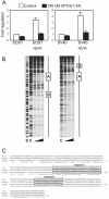
FIG. 1
FIG. 1
PTH responsiveness and transcription factor binding sites in the SOST bone enhancer. (A) Analysis of SOST promoter and bone enhancer regulation by PTH using reporter gene assays. UMR-106 cells were transfected with reporter plasmids containing 2 kb of the SOST proximal promoter (left) or the SV40 promoter (right) with or without upstream SOST bone enhancer. Eight hours after transfection, cells were stimulated for 16 h with 100 nM PTH(1-34) (filled bars) or solvent control (open bars). Subsequently, luciferase activity was determined. Shown are means and SE of relative luciferase activity from five independent experiments. (B) Mapping of transcription factor binding sites using DNase I footprint analysis. DNA fragments comprising the human SOST bone enhancer were either radiolabeled on the forward (left gel) or reverse strand (right gel) and were incubated with increasing amounts of UMR-106 cell nuclear extract (lanes 3–5). Lane C, control digestion without nuclear extract; lane S, dideoxy cytidine sequencing reaction control lane. (C) Nucleotide sequence of the SOST bone enhancer region from mouse, rat, and human. *Sequence identity. Footprint regions A and B are boxed.
FIG. 2
FIG. 2
Mutation analysis of the SOST bone enhancer. Deletion and point mutation constructs as depicted in the figure were tested for transcriptional activation in reporter gene assays using UMR-106 cells. Percent luciferase activity is expressed relative to the level of luciferase activity obtained with the intact SOST bone enhancer (p5). Values represent means and SE of five independent transfection experiments. The two protein binding regions A and B identified by footprint analysis are highlighted in gray.
FIG. 3
FIG. 3
The A2 element of the SOST bone enhancer binds MEF2 transcription factors. (A) Gel retardation assay using labeled double-stranded wildtype (WT) oligonucleotide comprising the MEF2 binding site and nuclear extract from UMR-106 cells. Competition experiments were performed with 50-fold molar excess of the same unlabeled oligonucleotide (WT) or mutated oligonucleotides as listed in B. The specific protein–DNA complex is indicated by an arrow. (B) Summary of the mutational analysis of the MEF2 binding site using gel retardation competition assays. The MEF2 consensus binding sequence is underlined. (C) Antibody-induced gel retardation supershift assays. Protein–DNA complexes as in A were incubated with two different amounts of a pan-specific MEF2 antibody or a p38 antibody as a control before gel electrophoresis. Protein–DNA complexes are indicated by a solid arrow and the additionally retarded antibody–protein–DNA complexes (supershifts) by an open arrow. (D) Analysis of SOST bone enhancer regulation by PTH using reporter gene assay. UMR-106 cells were transfected with reporter plasmids containing the SV40 promoter with or without upstream full-length SOST bone enhancer or only the A2 element (MEF2 TFBS). Eight hours after transfection, cells were stimulated for 16 h with 100 nM PTH(1-34) (filled bars) or solvent control (open bars). Subsequently, luciferase activity was determined. Shown are means and SE of relative activity of three independent experiments.
FIG. 4
FIG. 4
MEF2 RNA expression in bone tissues. MEF2A, B, C, and D mRNA expressions were determined by qPCR in adult rat femur and UMR-106 cells using specific TaqMan probes that detect all known isoforms for each MEF2 gene. For comparison, expression was also measured in heart, brain, and liver. Relative expression levels are shown. Values represent means and SE of three independent experiments.
FIG. 5
FIG. 5
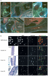
Localization of MEF2 RNA and protein in embryonic and adult bone. (A) In situ hybridization of MEF2C in mouse embryo at E16.5. MEF2C expression was detected by in situ hybridization with a fluorescently labeled MEF2C probe (red) and counterstaining with the nuclear stain DAPI. Sagittal sections show expression MEF2C expression in brain, nerve ganglia, and muscle, as well as in ossification sites such as ribs, hip bone, digits, and jaw. (B) Localization of MEF2 transcription factors and sclerostin in UMR-106 cells and osteocytes by immunohistochemistry. MEF2 (red) and sclerostin (green) were detected by double immunofluorescence labeling in permeabilized UMR-106 cells, in calvariae and femurs of 1 m-old (1m) mice, and in femurs of newborn (P0) mice. Insets show higher magnifications of the area indicated by a dotted box. Nuclear DAPI staining of UMR-106 cells and toluidine blue staining (T-blue) of representative bone sections are shown at lower magnification for orientation. Scale bars: 15 μm.
FIG. 6
FIG. 6
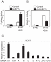
MEF2A, C, and D control SOST expression. (A) SOST bone enhancer activity is stimulated by MEF2. COS-7 cells were co-transfected with a human MEF2C expression plasmid or empty vector and reporter plasmids containing the 2-kb SOST promoter with or without upstream SOST bone enhancer. Subsequently, luciferase activity was determined. Shown are means and SE of relative luciferase activity from five independent experiments. (B) SOST bone enhancer activity is inhibited by a dominant-negative MEF2C mutant. UMR-106 cells were co-transfected with a dominant-negative human MEF2C expression plasmid dn(1–117)-hMEF2C or empty vector and reporter plasmids containing the SV40 promoter with or without the upstream SOST bone enhancer. Subsequently, luciferase activity was determined. Shown are means and SE of relative luciferase activity from five independent experiments. (C) Effect of MEF2A, B, C, and D expression inhibition on SOST expression. UMR-106 cells were transfected with specific siRNA against luciferase (Luc), SOST, MEF2A, B, C, and D, or combinations of MEF2s (A+C, A+D, C+D). Subsequently, SOST mRNA expression was determined by qPCR. Relative expression levels and SE are shown from four independent experiments.
FIG. 7
FIG. 7
Model of SOST gene regulation in osteocytes by PTH and MEF2 transcription factors. Expression of the osteocyte-specific bone formation inhibitor sclerostin (SOST) requires activation of the SOST bone enhancer by MEF2 transcription factors. Induction of bone formation by PTH-induced downregulation of SOST expression is mediated through MEF2 factors.

References

    1. Janssens K, Van Hul W. Molecular genetics of too much bone. Hum Mol Genet. 2002;11:2385–2393. - PubMed (VSports在线直播)
    1. van Bezooijen RL, ten Dijke P, Papapoulos SE, Lowik CW. SOST/sclerostin, an osteocyte-derived negative regulator of bone formation. Cytokine Growth Factor Rev. 2005;16:319–327. - "VSports" PubMed
    1. Balemans W, Ebeling M, Patel N, Van Hul E, Olson P, Dioszegi M, Lacza C, Wuyts W, Van Den EJ, Willems P, Paes-Alves AF, Hill S, Bueno M, Ramos FJ, Tacconi P, Dikkers FG, Stratakis C, Lindpaintner K, Vickery B, Foernzler D, Van Hul W. Increased bone density in sclerosteosis is due to the deficiency of a novel secreted protein (SOST) Hum Mol Genet. 2001;10:537–543. - PubMed
    1. Balemans W, Patel N, Ebeling M, Van Hul E, Wuyts W, Lacza C, Dioszegi M, Dikkers FG, Hildering P, Willems PJ, Verheij JB, Lindpaintner K, Vickery B, Foernzler D, Van Hul W. Identification of a 52 kb deletion downstream of the SOST gene in patients with van Buchem disease. J Med Genet. 2002;39:91–97. - PMC - PubMed
    1. Brunkow ME, Gardner JC, Van Ness J, Paeper BW, Kovacevich BR, Proll S, Skonier JE, Zhao L, Sabo PJ, Fu Y, Alisch RS, Gillett L, Colbert T, Tacconi P, Galas D, Hamersma H, Beighton P, Mulligan J. Bone dysplasia sclerosteosis results from loss of the SOST gene product, a novel cystine knotcontaining protein. Am J Hum Genet. 2001;68:577–589. - PMC - PubMed

"VSports注册入口" Publication types

MeSH terms (V体育官网入口)

Substances