Skip to main page content
U.S. flag

An official website of the United States government

Dot gov

The . gov means it’s official. Federal government websites often end in . gov or . mil. Before sharing sensitive information, make sure you’re on a federal government site. VSports app下载.

Https

The site is secure V体育官网. The https:// ensures that you are connecting to the official website and that any information you provide is encrypted and transmitted securely. .

. 2007 Jul 10;104(28):11609-14.
doi: 10.1073/pnas.0704898104. Epub 2007 Jul 2.

"VSports手机版" Thioredoxin is required for S-nitrosation of procaspase-3 and the inhibition of apoptosis in Jurkat cells

Affiliations

"VSports" Thioredoxin is required for S-nitrosation of procaspase-3 and the inhibition of apoptosis in Jurkat cells

Douglas A Mitchell et al. Proc Natl Acad Sci U S A. .

Abstract

S-nitrosation is a posttranslational, oxidative addition of NO to cysteine residues of proteins that has been proposed as a cGMP-independent signaling pathway [Hess DT, Matsumoto A, Kim SO, Marshall HE, Stamler JS (2005) Nat Rev Mol Cell Biol 6:150-166]. A paradox of S-nitrosation is that only a small set of reactive cysteines are modified in vivo despite the promiscuous reactivity NO exhibits with thiols, precluding the reaction of free NO as the primary mechanism of S-nitrosation. Here we show that a specific transnitrosation reaction between procaspase-3 and thioredoxin-1 (Trx) occurs in cultured human T cells and prevents apoptosis. Trx participation in catalyzing transnitrosation reactions in cells may be general because this protein has numerous protein-protein interactions and plays a key role in cellular redox homeostasis [Powis G, Montfort WR (2001) Annu Rev Pharmacol Toxicol 41:261-295], nitrosothiol content in cells [Haendeler J, Hoffmann J, Tischler V, Berk BC, Zeiher AM, Dimmeler S (2002) Nat Cell Biol 4:743-749], and antiapoptotic signaling VSports手机版. .


Conflict of interest statement

The authors declare no conflict of interest.

Figures

Fig. 1.
Fig. 1.
Trx transnitrosates procaspase-3 in Jurkat cells. (A) Trx is S-nitrosated in stimulated Jurkat cells. The Bradford assay was used to ensure equal protein loading. Trxred (C32/C35) and Trxox are visible. Lane 1, no treatment; lane 2, FasL; lane 3, etoposide; lane 4, IFN-γ. (B) Procaspase-3 is S-nitrosated under basal conditions; the method was the same as in A but using anti-caspase-3. Lane 1, no treatment; lane 2, etoposide; lane 3, IFN-γ. (C) Trx is S-nitrosated on C73 in Jurkat cells. Cells were stably transfected with V5-tagged Trx before immunoprecipitation using anti-V5 and the biotin switch. Lane 1, V5-Trx-WT; lane 2, V5-Trx-E70A/K72A (loss of contact); lane 3, V5-Trx-C73S (dominant negative); lanes 4–6, same order as lanes 1–3. + and − indicate etoposide (etop.) treatment. (D) Trx is involved in the formation of procaspase-3–SNO. Jurkat cells were stably transfected with V5-Trx mutants before knockdown of endogenous Trx using shRNA (3′ UTR). Densitometric analysis was performed on blots probed with HRP-conjugated neutravidin (NA-HRP) and anti-caspase-3 after an anti-caspase-3 immunoprecipitation followed by the biotin switch method. Black bars, nontransfected Jurkat cells; blue bars, cells transfected with shRNA; gray bars, cells transfected with V5-Trx-WT and shRNA; red bars, V5-Trx-C73S and shRNA; purple bars, V5-Trx-E70A/K72A and shRNA. + and − indicate etoposide (etop.) treatment. Asterisks indicate statistical significance between samples that were untreated (∗) and treated with etoposide (∗∗). P value was calculated by ANOVA. (E) Same as in D, but lysate was tested for AcDEVD-ase activity. Values were normalized to a sample that did not receive etoposide. The color scheme identical to that of D. Asterisks indicate significance between Jurkat cells that were not transfected with V5-Trx (∗, Student's t test) and cells that received a V5-Trx construct in addition to the shRNA plasmid (∗∗, ANOVA).
Fig. 2.
Fig. 2.
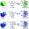
Model for nitrosothiol transfer between Trx and caspase-3. After apoptotic stimulation, endogenous WT Trx binds to and transfers +NO (blue and red spheres) to caspase-3, which leads to inhibition of apoptosis (healthy cell is shown in green). Cells transfected with a dominant negative (D.N.) or loss-of-contact (L.C.) Trx mutant do not inhibit apoptosis (apoptotic cell is shown in blue). Residues of Trx involved in caspase binding and transfer are shown in stick. PyMol and Protein Data Bank entries 1ERT and 1NME were used to construct this figure.
Fig. 3.
Fig. 3.
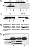
Trx and (pro)caspase-3 interact. (A) Anti-caspase-3 Western blot from Ni-NTA magnetocapture assay. WT His-Trx purifies caspase-3 from apoptotic Jurkat lysate. Lane 1, nontreated; lane 2, TNF-α; lane 3, staurosporine (His-Trx-E70A/K72A does not purify caspase-3); lanes 4–6, same as WT; lane 7, no His-Trx loaded (n.l.). (B) Coomassie-stained gel from an experiment identical to that in A. Lanes 1–3, same treatment as in A; lanes 4–6, same treatment as in A but using His-Trx-C69S as bait. (C) Sequence of His-Trx. Caspase-3 cut site is highlighted. (D) Coomassie-stained gel of caspase-3 proteolysis assay. Leftmost lane, molecular weight marker; lane 1, Trx-WT; lane 2, Trx-C69S; lane 3, Trx-D64A.
Fig. 4.
Fig. 4.
Urea denaturation of Trx proteins. Trx-C69S (open circles) has a weaker fold relative to WT (filled squares) and Trx-C73S (filled circles). Differences in the baseline CD signals, visible at lower urea concentration, were due to variance in the protein concentration (confirmed by the BCA method, quantitative amino acid analysis, and dry weight). Measurement of ΔGfold° is independent of protein concentration. Intrinsic tryptophan fluorescence gave similar results (data not shown).

"V体育平台登录" References

    1. Denninger JW, Marletta MA. Biochim Biophys Acta. 1999;1411:334–350. - PubMed
    1. Hess DT, Matsumoto A, Kim SO, Marshall HE, Stamler JS. Nat Rev Mol Cell Biol. 2005;6:150–166. - "V体育ios版" PubMed
    1. Foster MW, McMahon TJ, Stamler JS. Trends Mol Med. 2003;9:160–168. - PubMed
    1. Mannick JB, Hausladen A, Liu L, Hess DT, Zeng M, Miao QX, Kane LS, Gow AJ, Stamler JS. Science. 1999;284:651–654. - PubMed
    1. Mannick JB, Miao XQ, Stamler JS. J Biol Chem. 1997;272:24125–24128. - PubMed (V体育官网)

Publication types