Skip to main page content
U.S. flag

An official website of the United States government

Dot gov

The . gov means it’s official. Federal government websites often end in . gov or . mil. Before sharing sensitive information, make sure you’re on a federal government site VSports app下载. .

Https

The site is secure V体育官网. The https:// ensures that you are connecting to the official website and that any information you provide is encrypted and transmitted securely. .

Comparative Study
. 2007 Jun;11(6):539-54.
doi: 10.1016/j.ccr.2007.04.017.

Genes that distinguish physiological and pathological angiogenesis

Affiliations
Comparative Study

Genes that distinguish physiological and pathological angiogenesis

Steven Seaman et al. Cancer Cell. 2007 Jun.

Abstract

To unravel the normal vasculature transcriptome and determine how it is altered by neighboring malignant cells, we compared gene expression patterns of endothelial cells derived from the blood vessels of eight normal resting tissues, five tumors, and regenerating liver. Organ-specific endothelial genes were readily identified, including 27 from brain. We also identified 25 transcripts overexpressed in tumor versus normal endothelium, including 13 that were not found in the angiogenic endothelium of regenerating liver VSports手机版. Most of the shared angiogenesis genes have expected roles in cell-cycle control, but those specific for tumor endothelium were primarily cell surface molecules of uncertain function. These studies reveal striking differences between physiological and pathological angiogenesis potentially important for the development of tumor-specific, vascular-targeted therapies. .


Figures

Figure 1
Figure 1. Purification of ECs from normal and malignant tissues
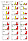
A: Immunofluorescence staining of heart tissue demonstrated co-localization of CD105 (green) with VE-cadherin (red) in the vessels. Scale bar, 20 μm. B: Immunofluorescence staining of liver tissue with CD105 (green). Scale bar, 20 μm. C: A QPCR analysis was used to assess the purity of the EC preparations. QPCR analysis was performed on cDNA generated directly from unfractionated normal whole tissues (WT) or from purified ECs isolated from normal tissues (N-ECs) or the tumors (T-ECs) indicated. The endothelial-specific transcript VE-cadherin was enriched 110 to 530-fold in the endothelial fractions. The modest level of VE-cadherin found in the unfractionated heart and lung sample is presumably due to a higher proportion of ECs in these tissues. In this experiment, gene expression was normalized to that of the Eif4h, a gene found to be uniformly expressed in all cells as assessed by SAGE (Velculescu et al., 1999). Unfractionated brain was used to calibrate relative expression because this tissue had the lowest VE-cadherin expression levels. D: Model used to identify genes expressed during pathological but not physiological angiogenesis. ECs were isolated from normal resting livers, regenerating livers, or tumor bearing livers.
Figure 2
Figure 2. Gene expression in resting normal ECs, regenerating liver ECs and tumor ECs
A: Expression of AEMs is upregulated in regenerating liver ECs. Note the high expression levels of integrinβ3 and inhibin beta B only 6 hours following partial hepatectomy. For comparison, expression patterns of a BEM (Organic-anion-transporter 2; Oatp2) are also shown in the upper left panel. B: Expression of TEMs is upregulated in tumor ECs. Note the low basal expression of these genes in regenerating liver ECs similar to that observed in ECs of normal resting tissues. Gene expression was evaluated by real-time QPCR and compared with that of Srnp70, a gene found to be expressed at nearly identical levels in all ECs by SAGE. For AEMs and TEMs, the results are expressed as a ratio between the gene of interest and Srnp70 and are normalized to the average expression of all non-angiogenic normal ECs. For Oatp2, samples were normalized to the average expression in intestinal, heart and kidney ECs. For comparison, normal ECs from resting liver (time=0h) were grouped with the regenerating liver ECs.
Figure 3
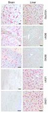
Figure 3. LEM and BEM genes identified by SAGE are expressed by ECs in vivo
Localization of mRNA in ECs (red stain) is demonstrated for the brain endothelial markers GLUT-1 (BEM1) and organic anion transporter 2 (BEM2), and the liver endothelial markers deoxyribonuclease 1-like 3 (LEM1) and oncogene induced transcript 3 (LEM2). Note that the BEMs are selectively expressed in brain endothelium whereas the LEMs are selectively expressed in liver endothelium. The endothelial control probe, VEGFR2, stains both brain and liver endothelium. Staining of LEMs is most prominent in the sinusoidal endothelium, wherein the nuclear body appears to stain most intensely. A dilute counterstain was applied to the sections to highlight the lack of detectable expression in the non-ECs of the tumors. Scale bars, 50 μM
Figure 4
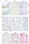
Figure 4. TEM genes identified by SAGE are expressed by tumor ECs in vivo
A: Localization of mRNA in tumor ECs (red stain) was demonstrated by examining Apelin and Doppel in subcutaneous implanted LLC tumors. Note the lack of detectable expression in the normal brain and liver tissues of these representative TEMs. B: Localization of various TEMs in the tumor endothelium of mice. Depicted are CD137 in a KM12SM tumor from the liver, CD109 and MiRP2 in SW620 subcutaneous tumors and CD276, PTPRN, ETSvg4 and Vscp in HCT116 subcutaneous tumors. C: CD276 mRNA is expressed in the vessels of human colorectal cancer. In situ hybridization revealed that CD276 mRNA is expressed predominantly in the vessels of human colorectal cancer (middle panel) with a pattern of staining similar to that of the control endothelial marker VEGFR2 (left panel). Note that in the case of CD276 the tumor cells also display positive staining, albeit less intense. At the margin between tumor (T) tissue and normal (N) colonic mucosa CD276 staining abruptly ends (right panel). The red extracellular staining around the normal crypts represents non-specific binding of the in situ hybridization reagents to the mucous (right panel) and is also present in control sections (data not shown). A dilute blue counterstain was applied to each of the sections. Scale bars, 50 μM
Figure 5
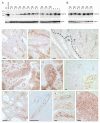
Figure 5. CD276 protein is overexpressed in human tumors
A: Immunoblotting with a CD276 monoclonal antibody revealed an upregulation of CD276 protein in colorectal tumors (T) compared to normal (N) colonic mucosa. Ten of the paired samples represent matched tissues taken from the same patient (P1-P10). CD276 protein migrates at a size similar to that observed in 293 cells transfected with the 4IgG-containing form of CD276 (293/CD276). The faint product present in 293 parent cells presumably represents low-level endogenous CD276 expression which can also be detected at the mRNA level in these cells by RT-PCR (data not shown). B: Immunoblotting with a CD276 monoclonal antibody revealed an upregulation of CD276 protein in lung tumors (T) compared to normal (N) adjacent lung tissue. The normal tissues in A and B were classified as normal based on gross morphology, but microscopic disease or inflammatory host cells may have contributed to the low level CD276 expression observed in these tissues. C-L: Immunohistochemical staining with a polyclonal CD276 antibody revealed a vessel-like pattern (brown stain) in colorectal cancer (C-E), non-small cell lung cancer (F-H), esophageal cancer (I-J), bladder cancer (K) and breast cancer (L). At the tumor margin (E) CD276 staining was weak or undetectable in normal colonic mucosa (N) but strong in the vessels of the adjacent tumor region (T). Vessels from normal tissues that failed to stain for CD276 were immunoreactive on control serial sections stained for endothelial proteins such as vWF (data not shown). In some tumors, the vessels appeared to stain most prominently (C-E and H-K) whereas in others, both tumor cells and tumor vessels were strongly positive (F-G and L). Note the strong cell surface staining pattern in the tumor epithelium under high power magnification (G). Many of the blood vessels were readily identified by the presence of blood cells in the lumen; for example see inset displaying higher power magnification of boxed region in (H). Sections were lightly counterstained with hematoxylin (blue stain). Scale bar, 50 μM.
Figure 6
Figure 6. Immunofluorescence staining reveals co-localization of CD276 with vWF in human colon cancer
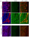
A: CD276 (green) was expressed predominantly by the tumor vessels of the colorectal cancer, but was also expressed at a lower level by the tumor cells themselves. Expression of CD276 in normal colonic mucosa was undetectable (top middle panel). As a control, vessels were stained for vWF (Red) which co-localized with CD276 only in the tumor sample. Scale bar, 100 μm. B: CD276 expression was undetectable in the angiogenic vessels of the developing corpus luteum. Scale bar, 200 μm. Sections were counterstained with DAPI (blue) which is shown in the left panels to highlight the epithelial cells.

Comment in (VSports手机版)

References

    1. Bradshaw HD., Jr. Molecular cloning and cell cycle-specific regulation of a functional human thymidine kinase gene. Proc Natl Acad Sci U S A. 1983;80:5588–5591. - "V体育ios版" PMC - PubMed
    1. Broll K, Richter G, Pauly S, Hofstaedter F, Schwarz H. CD137 expression in tumor vessel walls. High correlation with malignant tumors. Am J Clin Pathol. 2001;115:543–549. - PubMed
    1. Brooks PC, Clark RA, Cheresh DA. Requirement of vascular integrin alpha v beta 3 for angiogenesis. Science. 1994;264:569–571. - VSports注册入口 - PubMed
    1. Carmeliet P, Moons L, Luttun A, Vincenti V, Compernolle V, De Mol M, Wu Y, Bono F, Devy L, Beck H, et al. Synergism between vascular endothelial growth factor and placental growth factor contributes to angiogenesis and plasma extravasation in pathological conditions. Nat Med. 2001;7:575–583. - PubMed
    1. Carson-Walter EB, Watkins DN, Nanda A, Vogelstein B, Kinzler KW, St Croix B. Cell surface tumor endothelial markers are conserved in mice and humans. Cancer Res. 2001;61:6649–6655. - PubMed

Publication types

MeSH terms