Skip to main page content (VSports app下载)
U.S. flag

An official website of the United States government

Dot gov

The . gov means it’s official. Federal government websites often end in . gov or VSports app下载. mil. Before sharing sensitive information, make sure you’re on a federal government site. .

Https

The site is secure V体育官网. The https:// ensures that you are connecting to the official website and that any information you provide is encrypted and transmitted securely. .

. 2007 Mar 15;67(6):2657-65.
doi: 10.1158/0008-5472.CAN-06-1997.

Molecular cloning of hMena (ENAH) and its splice variant hMena+11a: epidermal growth factor increases their expression and stimulates hMena+11a phosphorylation in breast cancer cell lines (VSports)

Affiliations

Molecular cloning of hMena (ENAH) and its splice variant hMena+11a: epidermal growth factor increases their expression and stimulates hMena+11a phosphorylation in breast cancer cell lines (V体育ios版)

"V体育官网入口" Francesca Di Modugno et al. Cancer Res. .

Abstract

hMena (ENAH), an actin regulatory protein involved in the control of cell motility and adhesion, is modulated during human breast carcinogenesis. In fact, whereas undetectable in normal mammary epithelium, hMena becomes overexpressed in high-risk benign lesions and primary and metastatic tumors. In vivo, hMena overexpression correlates with the HER-2(+)/ER(-)/Ki67(+) unfavorable prognostic phenotype. In vitro, neuregulin-1 up-regulates whereas Herceptin treatment down-modulates hMena expression, suggesting that it may couple tyrosine kinase receptor signaling to the actin cytoskeleton. Herein, we report the cloning of hMena and of a splice variant, hMena(+11a), which contains an additional exon corresponding to 21 amino acids located in the EVH2 domain, from a breast carcinoma cell line of epithelial phenotype. Whereas hMena overexpression consistently characterizes the transformed phenotype of tumor cells of different lineages, hMena(+11a) isoform is concomitantly present only in epithelial tumor cell lines. In breast cancer cell lines, epidermal growth factor (EGF) treatment promotes concomitant up-regulation of hMena and hMena(+11a), resulting in an increase of the fraction of phosphorylated hMena(+11a) isoform only. hMena(+11a) overexpression and phosphorylation leads to increased p42/44 mitogen-activated protein kinase (MAPK) activation and cell proliferation as evidenced in hMena(+11a)-transfected breast cancer cell lines. On the contrary, hMena knockdown induces reduction of p42/44 MAPK phosphorylation and of the proliferative response to EGF. The present data provide new insight into the relevance of actin cytoskeleton regulatory proteins and, in particular, of hMena isoforms in coupling multiple signaling pathways involved in breast cancer. VSports手机版.


Figures (VSports注册入口)

Figure 1
Figure 1
Analysis of hMena transcripts. A, hMena amplified product by RT-PCR in SBT breast cancer cell line analyzed by agarose gel electrophoresis and ethidium bromide staining. B, PCR done with primers (MTC1f and MTC4r) flanking the region of alternative splicing on the pcDNA3.1 vectors containing the hMena sequences. PCR products were analyzed by agarose gel electrophoresis and ethidium bromide staining. C, diagrammatic representation of the two hMena gene transcripts. Exons are enumerated and the relative protein domains are indicated. D, amino acid sequence of the exon 11a coded peptide. Arrows, predicted phosphorylation residues.
Figure 2
Figure 2
hMena and hMena+11a isoform characterization and expression in tumor cell lines. A, analysis of the proteins encoded by hMena and hMena+11a. In vitro translated hMena and hMena+11a analyzed by Western blot analysis with the CKLK1 antibody (10 μg/mL). Protein extracts (30 μg) of SBT breast cancer cell line (used to obtain hMena+11a and hMena cDNAs) were also tested by Western blot to identify the corresponding in vitro translated protein bands in whole-cell lysates. B, protein extracts from cells derived from breast tumor cell lines (MDAMB361, SBT, DAL, T47D, SKBr3, MCF7, and BT474) and mammary gland tissue (Clontech), lung cancer cell lines (Calu3 and A427), cervix tumor cell lines (SiHa and CaSki), normal human keratinocytes (NHK), colon cancer cell lines (LS180 and HT29), and cecum tissue (Clontech), astrocytoma (ADF), and glioma cell lines (U251, U87MG, U373, and T98G) were immunoblotted with anti-hMena CKLK1 antibody. As loading control, the same blots were probed with anti-actin monoclonal antibody. C, analysis of hMena splice variants expression by RT-PCR. RT-PCR done with primers flanking the region of alternative splicing (MTC1f and MTC4r) on mRNA samples of epithelial breast cancer (MCF7 and SBT), melanoma (ME10538 and MAS), and glioma (T98G) cell lines. cDNAs of the pcDNA3.1 vectors containing the hMena and hMena+11a sequences were also amplified with the same primers. PCR products were analyzed by agarose gel electrophoresis and ethidium bromide staining. D, analysis of hMena expression by two-dimensional electrophoresis. Proteins from two different breast carcinoma cell lines (SBT and MCF7), a colon cancer cell line (LS180), and a glioma cell line (T98G) were resolved by pI in the first dimension on a pH 3 to 10 nonlinear range and by relative molecular mass in the second dimension on a 10% acrylamide SDS-PAGE. Proteins were then electrotransferred to nitrocellulose and hMena reactivity was revealed by Western blot (WB). Protein spot sets were compared by using Progenesis PG240 v2005 software (Nonlinear Dynamics). The range of pI values assigned to detected spots is indicated. Two sets of spots with different molecular mass and pI ranging between 5.4 to 6.0 (lower protein spots) and 5.8 to 6.2 (upper protein spots), indicated as hMena and hMena11a, respectively, are clearly discriminated.
Figure 3
Figure 3
hMena expression is increased by EGF treatment in epithelial breast cancer cell lines. A, MDAMB361 cells were serum starved (24 h) and stimulated for 24 h with the indicated amount of EGF. Lysates (30 μg) were analyzed by Western blot with anti-hMena and anti-actin antibodies. B, evaluation of hMena expression by Western blot in T47D breast tumor cells untreated and treated for 24, 48, and 72 h with 75 ng/mL of EGF, or after removal of EGF at 24 and 48 h after 24-h treatment (withdrawal). C, T47D cells untreated, treated with EGF for 24 h, or treated with EGF for 24 h after 30-min incubation with the EGFR inhibitor AG1478 (500 nmol/L) were evaluated for hMena expression. D to F, fluorescence analysis of hMena labeling (green) on T47D cells untreated (D), treated with 75 ng/mL of EGF for 24 h (E), or treated with AG1478 and EGF for 24 h (F) after overnight serum starvation. Blue, overlay DAPI nuclear labeling.
Figure 4
Figure 4
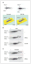
hMena and hMena+11a levels are enhanced and hMena+11a undergoes phosphorylation in response to EGF treatment. A, proteins from the T47D cell line either untreated or treated with EGF for 24 h were resolved by two-dimensional electrophoresis on a pH 3 to 10 nonlinear range and 7.5% acrylamide SDS-PAGE. Proteins were then electrontransferred to nitrocellulose and hMena reactivity was revealed by Western blot. Protein spot trains were compared by using Progenesis PG240 v2005 software (Nonlinear Dynamics). Colored panels are three-dimensional view densitometric analysis of the hMena spots. Arrows, protein spots affected by EGF treatment. B, proteins from the T47D cell line were obtained from cells treated with EGF alone or after pretreatment with EGFR inhibitor AG1478. Lysates from EGF-treated cells were incubated with or without λ-PPase. Proteins were resolved by two-dimensional electrophoresis on a pH 3 to 10 nonlinear range and 7.5% acrylamide SDS-PAGE. Proteins were processed as described in (A). Arrows, protein spots affected by treatments. C, lysates from the untreated T47D cell line were incubated with or without λ-PPase, resolved by two-dimensional electrophoresis, and transferred for Western blot as in (B). Arrows, hMena+11a spots after phosphatase treatment, indicating that a fraction of hMena+11a is constitutively phosphorylated in untreated cells.
Figure 5
Figure 5
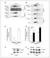
hMena+11a phosphorylation, proliferation rate, and p42/44 MAPK activity are enhanced by the transfection of hMena+11a in MCF7 and SKBr3 breast cancer cell lines. A, proteins from the MCF7 cell line either transfected with hMena+11a or empty vector (pcDNA3) were incubated without or with λ-PPase and resolved by two-dimensional electrophoresis on a pH 3 to 10 nonlinear range and 7.5% acrylamide SDS-PAGE. Proteins were then electrontransferred to nitrocellulose and hMena reactivity was revealed by Western blot. Protein spot trains were compared by using Progenesis PG240 v2005 software (Nonlinear Dynamics). The set of spots corresponding to hMena and hMena+11a is indicated. The phosphorylation threshold (arrows and bar) has been arbitrarily defined on the pattern of EGF- and λ-PPase–treated samples, these two conditions being considered the putative maximal and minimal rates of phosphorylation, respectively. B, proteins from the SKBr3 cell line transfected with either hMena+11a or empty vector were incubated with or without EGF and analyzed as described for MCF7 in (A). C, the proliferation rate is increased in hMena+11a–transfected MCF7 cells. MCF7/hMena+11a and MCF7/pcDNA3 cells were seeded in sextuplicate in 96-well plates (1 × 10⁴ per well) and cell proliferation was assessed after 72 h by measuring [3H]thymidine incorporation as described in Materials and Methods. Columns, mean of three different experiments; bars, SD. **, P < 0.05. D, SKBr3 cells were seeded in sextuplicate in 96-well plates (1 × 10⁴ per well). After 24 h, cells were transient transfected, cultured for 48 h, and treated with EGF (75 ng/mL) for 24 h. Cell proliferation was assessed as described above. E and F, the phosphorylation status of MAPK (p42/44) from whole-cell lysates (50 μg) of cells was assessed by Western blot with phosphospecific antibodies. Densitometric quantitation of anti–phosphorylated p42/44 MAPK immunoreactivity was determined by Quantity One software (Bio-Rad) and normalized in comparison with the total MAPK reactivity. Values are indicated at the bottom. Membranes were sequentially stripped and reprobed with the indicated total and phosphospecific antibodies.
Figure 6
Figure 6
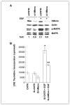
hMena knockdown reduces EGF-mediated MAPK phosphorylation and cell proliferation in BT474 breast cancer cell line. A, Western blot analysis of BT474 breast cancer cell line after 72-h transfection with control and hMena-specific siRNAs, untreated or EGF treated (75 ng/mL for 24 h). Silencing of hMena reduces the EGF-induced MAPK (p42/44) phosphorylation. The hMena expression and phosphorylation status of MAPK (p42/44) from whole-cell lysates were assessed. Densitometric quantitation of anti–phoshorylated p42/44 MAPK immunoreactivity was determined by Quantity One software (Bio-Rad) and normalized in comparison with the total MAPK reactivity. Values are indicated at the bottom. Membranes were sequentially stripped and reprobed with the indicated total and phosphospecific antibodies. B, silencing of hMena reduces EGF-mediated cell proliferation of BT474 cell line. Proliferation assays were conducted 72 h after the siRNA transfection by measuring [3H]thymidine incorporation as described in Materials and Methods. CNTR, untransfected BT474 cells; si-CNTR, BT474 cells transfected with nontargeting siRNA; si-hMena, BT474 cells transfected with specific hMena siRNA SMART pool. Columns, mean of three different experiments; bars, SD. *, P < 0.05, compared with si-CNTR. **, P < 0.05, compared with EGF (stimulated cells).

References

    1. Wulfkuhle JD, Sgroi DC, Krutzsch H, et al. Proteomics of human breast ductal carcinoma in situ. Cancer Res. 2002;62:6740–9. - V体育ios版 - PubMed
    1. Wang W, Goswami S, Lapidus K, et al. Identification and testing of a gene expression signature of invasive carcinoma cells within primary mammary tumors. Cancer Res. 2004;64:8585–94. - VSports手机版 - PubMed
    1. Di Modugno F, Bronzi G, Scanlan MJ, et al. Human Mena protein, a serex-defined antigen overexpressed in breast cancer eliciting both humoral and CD8+ T-cell immune response. Int J Cancer. 2004;109:909–18. - "VSports最新版本" PubMed
    1. Di Modugno F, Mottolese M, Di Benedetto A, et al. The cytoskeleton regulatory protein hmena (ENAH) is over-expressed in human benign breast lesions with high risk of transformation and human epidermal growth factor receptor-2-positive/hormonal receptor-negative tumors. Clin Cancer Res. 2006;12:1470–8. - "VSports最新版本" PubMed
    1. Bear JE, Svitkina TM, Krause M, et al. Antagonism between Ena/VASP proteins and actin filament capping regulates fibroblast motility. Cell. 2002;109:509–21. - PubMed

Publication types

MeSH terms

Substances

LinkOut - more resources