Skip to main page content
U.S. flag

An official website of the United States government

Dot gov

The . gov means it’s official. Federal government websites often end in . gov or . mil. Before sharing sensitive information, make sure you’re on a federal government site VSports app下载. .

Https

The site is secure. The https:// ensures that you are connecting to the official website and that any information you provide is encrypted and transmitted securely. V体育官网.

. 2007 Feb 26;176(5):709-18.
doi: 10.1083/jcb.200610046.

Critical role of the extracellular signal-regulated kinase-MAPK pathway in osteoblast differentiation and skeletal development (VSports在线直播)

Affiliations

"VSports注册入口" Critical role of the extracellular signal-regulated kinase-MAPK pathway in osteoblast differentiation and skeletal development

Chunxi Ge et al. J Cell Biol. .

Abstract (VSports注册入口)

The extracellular signal-regulated kinase (ERK)-mitogen-activated protein kinase (MAPK) pathway provides a major link between the cell surface and nucleus to control proliferation and differentiation. However, its in vivo role in skeletal development is unknown. A transgenic approach was used to establish a role for this pathway in bone. MAPK stimulation achieved by selective expression of constitutively active MAPK/ERK1 (MEK-SP) in osteoblasts accelerated in vitro differentiation of calvarial cells, as well as in vivo bone development, whereas dominant-negative MEK1 was inhibitory VSports手机版. The involvement of the RUNX2 transcription factor in this response was established in two ways: (a) RUNX2 phosphorylation and transcriptional activity were elevated in calvarial osteoblasts from TgMek-sp mice and reduced in cells from TgMek-dn mice, and (b) crossing TgMek-sp mice with Runx2+/- animals partially rescued the hypomorphic clavicles and undemineralized calvaria associated with Runx2 haploinsufficiency, whereas TgMek-dn; Runx2+/- mice had a more severe skeletal phenotype. This work establishes an important in vivo function for the ERK-MAPK pathway in bone that involves stimulation of RUNX2 phosphorylation and transcriptional activity. .


Figures

Figure 1.
Figure 1.
Development of transgenic mouse lines overexpressing constitutively active and dominant-negative MEK1 in osteoblasts. (a) Schematic representation of a transgene construct. A −647 to +13 bp fragment of the murine mOG2 promoter was used to drive expression of constitutively active (MEK-SP) and dominant-negative MEK1 mutants in osteoblasts. (b) Tissue distribution of transgene expression. Total RNA was isolated from the indicated tissues of 4-wk-old mice, and transgene expression measured by RT-PCR. The lines used, from left to right, were as follows: DN288, DN388, DN315, SP413, SP221, and SP211. GAPDH mRNA levels are shown as a control for RNA loading. Transgene expression was not detected in muscle, brain, or liver (not depicted). (c) Transgene expression time course during embryonic development. Results with SP221 and DN288 are shown. (d) Distribution of transgene expression in long bone. In situ hybridization was used to localize transgene mRNA to endosteal and select trabecular surfaces of a newborn TgMek-dn femur (line DN288). (top) Hematoxylin and eosin–stained section; middle, in situ hybridization with digoxigenin-labeled sense probe; bottom, antisense probe. Inset, 2× magnification of boxed area. Bar, 250 μm.
Figure 2.
Figure 2.
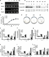
Altered osteoblast differentiation in calvarial cells from TgMek-dn and -sp mice. Cells were isolated from calvaria of newborn wild-type and transgenic animals. (a) Time course of transgene expression. Cells were plated and grown in differentiating medium for the indicated times before measurement of transgene mRNA by RT-PCR. (b) Mek-dn and -sp transgene expression alters ERK phosphorylation. Cells were grown as in a and harvested after 10 d for measurement of total and phospho-ERK by Western blotting. The indicated groups were treated with the Raf inhibitor ZM336372 2 h before harvest. (c) Transgene expression does not alter cell growth. (d–j) MEK-DN inhibits, whereas MEK-SP stimulates, osteoblast differentiation. The following differentiation markers were measured: mineralized nodules in 14-d cultures (d), alkaline phosphatase activity (e), total cell layer–associated, acid-extractable calcium (f), and OCN, BSP, ALP, and Runx2 mRNA levels (g–j; all measured by real-time RT-PCR). Values are the mean ± the SD of triplicate independent samples.
Figure 3.
Figure 3.
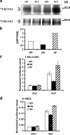
Changes in RUNX2 phosphorylation and transcriptional activity in osteoblasts from TgMek-dn and -sp mice. (a and b) Regulation of RUNX2 phosphorylation. Calvarial cells were grown under differentiating conditions for 7 d before metabolic labeling with [32P]orthophosphate or [35S]methionine/cysteine (to normalize for total RUNX2) and immunoprecipitation with an anti-Runx2 antibody. Each IP reaction contained 500 μg total protein. (b) Normalized 32P incorporation into RUNX2. (c and d) Runx2-dependent transcriptional activity. Cells were transfected with p1.3mOG2-luc (c) or p6OSE2mOG2-luc (d) plasmids and grown under differentiating conditions for the times indicated before measurement of luciferase activity. Values are the means ± the SD of triplicate independent samples.
Figure 4.
Figure 4.
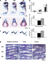
Altered skeletal development in TgMek-dn and -sp mice. (a) Whole mounts of E15.5 skeletons stained with alcian blue and alizarin red. (d) Effects of transgene expression on embryo weights. (b and e) Cranial bones showing differences in mineralization (b) and quantification of mineralized area (expressed as the percentage of total calvarial area; e). (c and f) Hindlimbs showing differences in the size of bones with transgene expression (c) and quantification of femur lengths (f). (g) Histology of long bones from wild-type, TgMek-dn, and -sp mice. Note delay in bony collar and trabecular bone in TgMek-dn embryos. Statistical analysis values are expressed as the mean ± the SD. n = 8 mice/group. *, significantly different from wild type at P < 0.01.
Figure 5.
Figure 5.
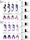
Genetic interactions between Mek-dn and -sp transgenes and Runx2. TgMek-dn or -sp mice were crossed with Runx2+/− mice to generate the genotypes indicated. (a–d) Partial rescue of CCD phenotype in Runx2+/− mice with Mek-sp. (a) Skeletal whole mounts of newborn mice stained with alcian blue and alizarin red (top), isolated clavicles (middle), and crania (bottom). (b–d) measurements of femur length (b), clavicle areas (c), and mineralized area of calvaria (expressed as a fraction of total calvarial area). (e–h) Increased severity of CCD phenotype with Mek-dn. Groups are as in a–d. Statistical analysis values are expressed as the means ± the SD. n = 8 mice/group. Comparisons are indicated by bars. *, significantly different at P < 0.01.

References

    1. Adams, M., M.J. Reginato, D. Shao, M.A. Lazar, and V.K. Chatterjee. 1997. Transcriptional activation by peroxisome proliferator-activated receptor gamma is inhibited by phosphorylation at a consensus mitogen-activated protein kinase site. J. Biol. Chem. 272:5128–5132. - PubMed
    1. Celil, A.B., and P.G. Campbell. 2005. BMP-2 and insulin-like growth factor-I mediate Osterix (Osx) expression in human mesenchymal stem cells via the MAPK and protein kinase D signaling pathways. J. Biol. Chem. 280:31353–31359. - PubMed
    1. Celil, A.B., J.O. Hollinger, and P.G. Campbell. 2005. Osx transcriptional regulation is mediated by additional pathways to BMP2/Smad signaling. J. Cell. Biochem. 95:518–528. - PubMed
    1. Chen, C., A.J. Koh, N.S. Datta, J. Zhang, E.T. Keller, G. Xiao, R.T. Franceschi, N.J. D'Silva, and L.K. McCauley. 2004. Impact of the mitogen-activated protein kinase pathway on parathyroid hormone-related protein actions in osteoblasts. J. Biol. Chem. 279:29121–29129. - "VSports" PubMed
    1. Ducy, P., and G. Karsenty. 1995. Two distinct osteoblast-specific cis-acting elements control expression of a mouse osteocalcin gene. Mol. Cell. Biol. 15:1858–1869. - "VSports手机版" PMC - PubMed

Publication types

VSports在线直播 - MeSH terms

"VSports手机版" Substances