Skip to main page content (VSports在线直播)
U.S. flag

An official website of the United States government

Dot gov

The VSports app下载. gov means it’s official. Federal government websites often end in . gov or . mil. Before sharing sensitive information, make sure you’re on a federal government site. .

Https

The site is secure. The https:// ensures that you are connecting to the official website and that any information you provide is encrypted and transmitted securely. V体育官网.

. 2007 Feb;11(2):175-89.
doi: 10.1016/j.ccr.2006.11.024.

p53-deficient cells rely on ATM- and ATR-mediated checkpoint signaling through the p38MAPK/MK2 pathway for survival after DNA damage

Affiliations

p53-deficient cells rely on ATM- and ATR-mediated checkpoint signaling through the p38MAPK/MK2 pathway for survival after DNA damage

H Christian Reinhardt et al. Cancer Cell. 2007 Feb.

Abstract

In response to DNA damage, eukaryotic cells activate ATM-Chk2 and/or ATR-Chk1 to arrest the cell cycle and initiate DNA repair. We show that, in the absence of p53, cells depend on a third cell-cycle checkpoint pathway involving p38MAPK/MK2 for cell-cycle arrest and survival after DNA damage VSports手机版. MK2 depletion in p53-deficient cells, but not in p53 wild-type cells, caused abrogation of the Cdc25A-mediated S phase checkpoint after cisplatin exposure and loss of the Cdc25B-mediated G2/M checkpoint following doxorubicin treatment, resulting in mitotic catastrophe and pronounced regression of murine tumors in vivo. We show that the Chk1 inhibitor UCN-01 also potently inhibits MK2, suggesting that its clinical efficacy results from the simultaneous disruption of two critical checkpoint pathways in p53-defective cells. .


Figures
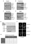
Figure 1
Figure 1. The p38MAPK/MK2pathway is activated by DNA- damaging drugs
(A–D) Kinetics of p38 MAPK and MK2 activation. U2OS cells were treated with 10μM cisplatin (A), 10μM camptothecin (B), 10μM doxorubicin (C) or DMSO control (D) for the indicated times. Cell lysates were probed for total and phosphorylated/activated forms of p38MAPK and MK2(MK-2) by Western blotting. β-actin served as a loading control. (E) MK2 activation is p38MAPK-dependent. U2OS cells were treated with the p38 MAPK specific inhibitor SB203580 (10μM) or DMSO vehicle for 30 min prior to exposure to chemotherapeutic drugs as in panels A–D. Total and phosphorylated/activated p38 was determined by immunoblotting as above. (F) Activation of MK2 parallels the formation of γH2AX nuclear foci. U2OS cells were either mock treated or incubated with cisplatin (10μM), camptothecin (10μM) or doxorubicin (10μM). Cells were immunostained 1 and 4h later using an antibody against γ-H2AX, and counterstained with DAPI. Scale bar, 2μm. (G) Summary of the requirement for ATM and/or ATR for the activation of MK2.
Figure 2
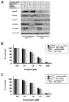
Figure 2. p53 deficient MEFs require MK2 for survival after DNA damage
(A) p21 induction is independent of MK2 activation. p53WT/WT and p53−/− MEFs stably expressing luciferase shRNA or MK2 shRNA were treated with 10μM cisplatin or 10μM doxorubicin. MK2activation and components of the p53 pathway were monitored by SDS-PAGE and Western blot analysis. (B, C) Clonogenic survival assay. p53WT/WT and p53−/− MEFs stably expressing luciferase shRNA or MK2 shRNA were mock treated or treated with increasing doses of cisplatin (B) or doxorubicin (C) for 4 hours, washed in PBS, trypsinized and replated at 5000 cells/10cm dish. 14 days later, surviving colonies were fixed, stained with crystal violet, and counted. Assays were performed in triplicate for each condition and normalized to mock treated cells. Asterisks denote statistically significant differences (2-tailed Student’s t-test, p<0.04).
Figure 3
Figure 3. Depletion of MK2 sensitizes p53-deficient MEFs to the anti-proliferative effects of cisplatin and doxorubicin by inducing mitotic catastrophe
p53WT/WT and p53−/− MEFs stably expressing RNAi hairpins against luciferase (A, C) or MK2(B, D) were treated with low dose cisplatin (1.0μM) or doxorubicin (0.1μM) for 30hr, fixed and stained with antibodies against phospho-histone H3, γ-H2AX and cleaved caspase-3. Positive staining for γ-H2AX in combination with phospho-histone H3 and cleaved caspase-3 labeling is indicative of mitotic catastrophe, was only observed in MK2 depleted p53−/− cells (panel D, arrows). Arrowhead in that panel shows a γ-H2AX-positive cell that does not stain for either phospho-histone H3 or cleaved caspase-3. Scale bar, 5μm.
Figure 4
Figure 4. MK2 depletion enhances regression of established tumors after DNA damaging chemotherapy in a murine model
(A) H-Ras-V12 transformed p53 −/− MEFs (Ferbeyre et al., 2002) were infected with lentiviruses encoding U6 promoter-driven luciferase shRNA or MK2shRNA, and CMV promoter-driven GFP. 3d post-infection, GFP expressing cells were selected by FACS and cultured for an additional 3d. Efficiency of MK2knock-down in the entire GFP-positive population was then assessed by immunoblotting of total cell lysates. (B) Following subcutaneous injection of 106 shRNA-containing cells into the flanks of NCR nude outbred mice, tumor growth was measured every 2 days, starting at day 6 post injection. Arrow indicates the start of intraperitoneal administration of DMSO, 2 mg/kg cisplatin, or 4 mg/kg doxorubicin on day 12, given 3 times/week. In the absence of DNA-damaging chemotherapy, the MK2depleted tumors were statistically significantly larger than the control tumors at each time point beginning on day 13 (Student’s t-test, 2-tailed, p<0.02). In contrast, after cisplatin or doxorubicin treatment the MK2depleted tumors were statistically smaller than the control tumors beginning on days 21 and 23, respectively (p<0.02). (C) Upper panels: dorsal view of the tumors in situ 14 days after initiation of the indicated treatments, corresponding to 26 days after tumor cell implantation. Middle panels: corresponding fluorescence images. Lower panels: close-up view of the excised tumors. (D) Tumor weight was analyzed at the 26 day endpoint.
Figure 5
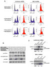
Figure 5. MAPKAP Kinase 2 mediates a G2/M arrest following doxorubicin treatment
(A) RNAi down-regulation of MK2 ablates the doxorubicin-induced G2/M checkpoint. p53−/− MEFs stably expressing control luciferase shRNA (left) or MK2 shRNA (right) were cultured in the absence or presence of 10 μM doxorubicin and cell cycle profiles analyzed 30h later by FACS using PI for DNA content (blue) and phospho-histone H3 staining as an indicator of mitosis (red). In the lower set of panels, nocodazole (100 ng/ml) was added 3h following doxorubicin addition. Note that in addition to loss of the prominent G2/M checkpoint, the G1 and S phase components are also eliminated in MK2-depleted cells following doxorubicin + nocodazole treatment. (B) Down-regulation of MK2 does not impair Chk1 activation. Luciferase shRNA-or MK2 shRNA expressing p53−/− MEFs were mock treated or exposed to 10μM doxorubicin or 10μM camptothecin for 30h. Total cell lysates were immunoblotted for the presence of MK2and total and activated forms of Chk1. (C) Doxorubicin and camptothecin-induced binding of Cdc25B to 14-3-3 is lost in MK2 depleted cells. p53−/− MEFs cells either expressing a luciferase hairpin (upper panel) or a MK2 specific hairpin (lower panel) were mock treated or treated with 10μM camptothecin (cam) or 10μM doxorubicin (dox) for 8 hr. The presence of 14-3-3-binding sites on Cdc25B was monitored by incubating the lysates with bead-bound GST-14-3-3β/ζ followed by immunoblotting of the pulled-down material.
Figure 6
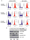
Figure 6. MK2 controls the S-phase checkpoint in response to cisplatin treatment
(A) RNAi down-regulation of MK2 ablates the cisplatin-induced S-phase checkpoint. p53−/− MEFs stably expressing control luciferase shRNA (left) or MK2shRNA (right) were cultured in the absence or presence of 10 μM cisplatin and cell cycle profiles analyzed 30 hr later by FACS using PI for DNA content (blue) and phospho-histone H3 staining as an indicator of mitosis (red). In the lower set of panels, nocodazole (100 ng/ml) was added 3 hrs following cisplatin addition. (B) Cisplatin-induced degradation of Cdc25A is impaired in MK2 depleted cells despite activation of Chk1. Luciferase shRNA- or MK2 shRNA expressing p53−/−MEFs were mock treated or exposed to 10μM cisplatin for 8 and 12 hr. Total cell lysates were immunoblotted for Cdc25A, total MK2 and activated Chk1. β-actin was used as a loading control.
Figure 7
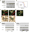
Figure 7. UCN-01 potently inhibits MK2 in vitro and in vivo
(A) MK2 is activated independently of Chk1. U2OS cells were either transfected with Chk1-specific siRNA oligonucleotides or GFP control oligonucleotides and MK2activation was monitored after treatment with cisplatin (10μM) or doxorubicin (10μM). Cell lysates were probed for total and phosphorylated/activated forms of MK2with an antibody detecting total Chk1, to monitor the knock down efficiency. (B) In vitro kinase assays were performed with Chk1 and MK2in the presence of increasing doses of UCN-01 using MK-2tide as a substrate. (C) Structural basis for UCN-01 inhibition of MK2. Ribbon diagrams (β-strands shaded yellow, α-helices shaded red, loops shaded green) and molecular surfaces of the Chk1:UCN-01 complex (panels 1, 4) and the MK2:staurosporine complex with UCN-01 modeled onto staurosporine (panels 2,3, 5). Panel 3 is rotated 90° from the view in panel 2; the staurosporine molecule is colored purple, UCN-01 is shown with carbon atoms colored cyan, oxygens red and nitrogens blue. The arrow points to the unique 7-hydroxy group of UCN-01 with Van der Waals radii indicated by dots. (D) UCN-01 inhibits MK2 in U2OS cells. Luciferase shRNA- or MAPKAP Kinase 2 shRNA expressing cells were incubated at 37°C or 42°C for 2 hr in the absence or presence of 250nM UCN-01. Cells were lysed and probed for total hsp-27, hsp-27 pS82, and MK2by immunoblotting. (E) UCN-01 inhibition of hsp-27 is independent of Chk1. GFP or Chk1 siRNA transfected U2OS cells were incubated at 42°C or 37°C for 2h in the absence or presence of 250nM UCN-01. Cells were then lysed and probed for total hsp-27, hsp-27 pS82, and Chk1 by immunoblotting.
Figure 8
Figure 8. A simplified model for re-wiring of cell cycle checkpoint pathways in p53-proficient and deficient cells
(A) Checkpoint function in p53-proficient cells is mediated primarily through a robust, sustained p53 response downstream of ATM, together with Chk1. Although not shown explicitly in the diagram, Chk1 also directly phosphorylates p53 (Shieh et al., 2000). Under these conditions the presence of MK2 is not required for cell survival after exposure to DNA damaging agents. (B) In p53-deficient cancer cells, checkpoint signaling following exposure to DNA damaging agents is mediated through the combined action of both the Chk1 and the p38 MAPK/MK2pathways. In this situation the p38MAPK/MK2 branch of checkpoint signaling becomes essential for cell survival after DNA damage. Both pathways are simultaneously inhibited by the indolocarbazole drug UCN-01.

References (V体育官网入口)

    1. Agner J, Falck J, Lukas J, Bartek J. Differential impact of diverse anticancer chemotherapeutics on the Cdc25A-degradation checkpoint pathway. Exp Cell Res. 2005;302:162–169. - PubMed
    1. Bartek J, Lukas J. Pathways governing G1/S transition and their response to DNA damage. FEBS Lett. 2001;490:117–122. - PubMed
    1. Bartek J, Lukas J. Chk1 and Chk2 kinases in checkpoint control and cancer. Cancer Cell. 2003;3:421–429. - PubMed
    1. Ben-Levy R, Leighton IA, Doza YN, Attwood P, Morrice N, Marshall CJ, Cohen P. Identification of novel phosphorylation sites required for activation of MAPKAP kinase-2. Embo J. 1995;14:5920–5930. - PMC - PubMed
    1. Block WD, Merkle D, Meek K, Lees-Miller SP. Selective inhibition of the DNA-dependent protein kinase (DNA-PK) by the radiosensitizing agent caffeine. Nucleic Acids Res. 2004;32:1967–1972. - PMC - PubMed

"VSports最新版本" Publication types

MeSH terms

Substances