<var date-time="gH5sCTx"></var>
Skip to main page content
U.S. flag

An official website of the United States government

Dot gov

The . gov means it’s official. Federal government websites often end in VSports app下载. gov or . mil. Before sharing sensitive information, make sure you’re on a federal government site. .

Https

The site is secure. The https:// ensures that you are connecting to the official website and that any information you provide is encrypted and transmitted securely. V体育官网.

. 2007 Jan;48(1):194-202.
doi: 10.1167/iovs.06-0632.

Magnetic resonance imaging evidence for widespread orbital dysinnervation in dominant Duane's retraction syndrome linked to the DURS2 locus

Affiliations

Magnetic resonance imaging evidence for widespread orbital dysinnervation in dominant Duane's retraction syndrome linked to the DURS2 locus

Joseph L Demer et al. Invest Ophthalmol Vis Sci. 2007 Jan.

Abstract

Purpose: High-resolution, multipositional magnetic resonance imaging (MRI) was used to demonstrate extraocular muscles (EOMs) and associated motor nerves in Duane retraction syndrome (DRS) linked to the DURS2 locus on chromosome 2 VSports手机版. .

Methods: Five male and three female affected members of two autosomal dominant DURS2 pedigrees were enrolled in the study. Coronal T(1)-weighted MRI of the orbits was obtained in multiple gaze positions, as well as with heavy T(2) weighting in the plane of the cranial nerves. MRI findings were correlated with motility. V体育安卓版.

Results: All subjects had unilateral or bilateral limitation of abduction, or of both abduction and adduction, with palpebral fissure narrowing and globe retraction in adduction. Orbital motor nerves were typically small, with the abducens nerve (cranial nerve [CN]6) often nondetectable. Lateral rectus (LR) muscles were structurally abnormal in seven subjects, with structural and motility evidence of oculomotor nerve (CN3) innervation from vertical rectus EOMs leading to A or V patterns of strabismus in three cases V体育ios版. Four cases had superior oblique, two cases superior rectus, and one case levator EOM hypoplasia. Only the medial and inferior rectus and inferior oblique EOMs were spared. Two cases had small CN3s. .

Conclusions: DRS linked to the DURS2 locus is associated with bilateral abnormalities of many orbital motor nerves, and structural abnormalities of all EOMs except those innervated by the inferior division of CN3. The LR may be coinnervated by CN3 branches normally destined for any other rectus EOMs. Therefore, DURS2-linked DRS is a diffuse congenital cranial dysinnervation disorder involving but not limited to CN6. VSports最新版本.


Figures

Figure 1
Figure 1
A-pattern in subject 4, showing esotropia in attempted upward gaze, and exotropia in central and downward gaze. Note limitation of vertical gaze, and left hypotropia. The upper eyelid configuration was created at surgery for blepharoptosis. There was prior strabismus surgery.
Figure 2
Figure 2
Quasicoronal MRI planes 2 mm thick of left orbit of subject 4 from pedigree SB, demonstrating a small, dysplastic SO, and dysplasia of the LPS, forming an abnormal slip extending toward the SO. The LR muscle was split into superior and inferior portions. The ON was small.
Figure 3
Figure 3
Quasicoronal MRI planes 2 mm thick of the right orbit of subject 4 from pedigree FY, demonstrating marked dysplasia of the LR, with continuity and apparent innervation of the LR by a branch of the inferior division of the oculomotor nerve (N3). The deep LR was also hypo-plastic. The SO was of normal size. The LPS was mildly dysplastic. Note the small ON cross section in the most anterior image at lower right.
Figure 4
Figure 4
Quasisagittal, T1-weighted MRI of subject 4 showing longitudinal splitting of the left LR into superior and inferior portions, with intercalated bright signal consistent with orbital fat: 312-μm resolution in 2-mm image plane. LG, orbital lobe of the lacrimal gland.
Figure 5
Figure 5
Oblique, axial, heavily T2-weighted MR images showing at left the normal course of the abducens nerve (CN6 dark) from the pons highlighted against the bright signal of the surrounding cerebrospinal fluid. In contrast, the CN6s of subject 4 with DURS2 appeared absent and the left CN6 of subject 9 was shown to be absent by the identical technique. In each panel, CN6 was demonstrated in the optimal 1-mm-thick image plane in which it appeared or was expected to appear based on surrounding landmarks.
Figure 6
Figure 6
Heavily T2-weighted axial MR images of 1-mm thickness at the level of the midbrain obtained in the plane of the optic chiasm. In all normal subjects, the oculomotor nerve (CN3) was prominent in multiple contiguous image planes. In subject 6 with DURS2, the right CN3 was hypoplastic.
Figure 7
Figure 7
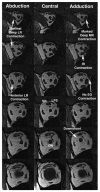
Coronal MRI of the right orbit of subject 1 with the clinical phenotype of unilateral DRS type 3 on the right. Image planes 2-mm thick spaced by 2 mm, resolution 312 μm in planes arranged from posterior at top to anterior at bottom. Fixation was by the clinically normal left eye. Note the minimal increase in the deep LR cross section on abduction, with a moderate increase in the anterior LR cross section on abduction. Although the deep MR exhibited a marked contractile increase in cross section, the eye adducted little, and the elevated position of the globe–ON junction indicated a downshoot. Note the increase in IR cross section in adduction suggestive of IR contraction, without corresponding increase in SO cross section. Note the normal positions of the MR and LR muscles near the globe–ON junction (bottom row), indicating normal stability of the horizontal rectus pulleys and ruling out sideslip as the cause of the downshoot in adduction.
Figure 8
Figure 8
Coronal MRI of left orbit in planes 2-mm thick spaced by 2 mm, resolution 234 μm in plane. Top row: subject 1 with clinical phenotype of DRS unilateral type 3 on the right only. Note hypoplasia of the deep portion of the LR, without a recognizable abducens nerve (CN6). The LR is also fissured. Also evident is the motor nerve to the MR. Bottom row: normal control subject, showing a normal LR cross section.

References

    1. Duane A. Congenital deficiency of abduction associated with impairment of adduction, retraction movements, contraction of the palpebral fissure and oblique movements of the eye. Arch Ophthalmol. 1905;34:133–159. - PubMed
    1. Huber A. Electrophysiology of the retraction syndromes. Br J Ophthalmol. 1974;58:293–300. - PMC - PubMed
    1. Strachan IM, Brown BH. Electromyography of extraocular muscles in Duane’s syndrome. Br J Ophthalmol. 1972;56:594–599. - PMC - PubMed
    1. Miller NR, Kiel SM, Green WR, Clark AW. Unilateral Duane’s retraction syndrome (type 1) Arch Ophthalmol. 1982;100:1468–1472. - PubMed
    1. Hotchkiss MG, Miller NR, Clark AW, Green WM. Bilateral Duane’s retraction syndrome: a clinical-pathologic case report. Arch Ophthalmol. 1980;98:870–874. - "VSports在线直播" PubMed

Publication types

MeSH terms