Skip to main page content
U.S. flag

An official website of the United States government

Dot gov

The . gov means it’s official. Federal government websites often end in . gov or . mil. Before sharing sensitive information, make sure you’re on a federal government site. VSports app下载.

Https

The site is secure. The https:// ensures that you are connecting to the official website and that any information you provide is encrypted and transmitted securely V体育官网. .

. 2007 Jan 5;100(1):70-8.
doi: 10.1161/01.RES.0000254788.47304.6e. Epub 2006 Dec 7.

"V体育ios版" Critical role of endothelial Notch1 signaling in postnatal angiogenesis

Affiliations

Critical role of endothelial Notch1 signaling in postnatal angiogenesis

Kyosuke Takeshita et al. Circ Res. .

Abstract

Notch receptors are important mediators of cell fate during embryogenesis, but their role in adult physiology, particularly in postnatal angiogenesis, remains unknown. Of the Notch receptors, only Notch1 and Notch4 are expressed in vascular endothelial cells VSports手机版. Here we show that blood flow recovery and postnatal neovascularization in response to hindlimb ischemia in haploinsufficient global or endothelial-specific Notch1(+/-) mice, but not Notch4(-/-) mice, were impaired compared with wild-type mice. The expression of vascular endothelial growth factor (VEGF) in response to ischemia was comparable between wild-type and Notch mutant mice, suggesting that Notch1 is downstream of VEGF signaling. Treatment of endothelial cells with VEGF increases presenilin proteolytic processing, gamma-secretase activity, Notch1 cleavage, and Hes-1 (hairy enhancer of split homolog-1) expression, all of which were blocked by treating endothelial cells with inhibitors of phosphatidylinositol 3-kinase/protein kinase Akt or infecting endothelial cells with a dominant-negative Akt mutant. Indeed, inhibition of gamma-secretase activity leads to decreased angiogenesis and inhibits VEGF-induced endothelial cell proliferation, migration, and survival. Overexpression of the active Notch1 intercellular domain rescued the inhibitory effects of gamma-secretase inhibitors on VEGF-induced angiogenesis. These findings indicate that the phosphatidylinositol 3-kinase/Akt pathway mediates gamma-secretase and Notch1 activation by VEGF and that Notch1 is critical for VEGF-induced postnatal angiogenesis. These results suggest that Notch1 may be a novel therapeutic target for improving angiogenic response and blood flow recovery in ischemic limbs. .


VSports注册入口 - Figures

Figure 1
Figure 1
Increase limb necrosis in Notch1 mutant mice. A, Expression of VEGF in control and ischemic limbs at 7 days was determined by immunohistochemical staining (top) and immunoblot analysis (bottom) of adductor muscle. Bar=25 μm. B, Photomicrographs of ischemic limb showing distal necrosis at 28 days (top, arrows). The percentage distribution of distal necrosis in WT, N1+/-, and ecN1+/- mice relative to the total number of mice in each group is shown (bottom). All mice survived hindlimb ischemia surgery and were analyzed. *P<0.05 compared with WT.
Figure 2
Figure 2
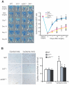
Impaired blood flow recovery and angiogenesis in Notch1 mutant mice. A, LBDF showing low-perfusion signal (dark blue) in ischemic hindlimbs of N1+/- and ecN1+/- mice, whereas high-perfusion signal (red to white) was observed in ischemic hindlimb of WT and N4-/- mice (left). Quantitative analysis of blood flow recovery following femoral artery ligation expressed as ischemic to control LDBF ratio in WT, N1+/-, ecN1+/-, and N4-/- mice (right). *P<0.0001, n=12 in each group. B, Isolectin B4 staining (brown-red color) of control (nonischemic) and ischemic adductor muscle tissues from hindlimbs of WT and Notch1 mutant mice (left). The photomicrographs are representative data from 8 individual mice from each group. Bar=25 μm. Quantitative analysis of capillary density (6 randomly selected fields from each slide; 3 slides per mice; 8 mice each) in WT and Notch1 mutant mice (number per hpf; ×400 magnification) (right). †P<0.001 compared with WT (nonischemic), *P<0.001 compared with WT (ischemic).
Figure 3
Figure 3
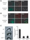
Impaired microvessel sprouting in Notch1 mutant mice. A, Immunofluorescence staining for Notch1 (green) and PECAM-1 (red) in skeletal muscle tissues from control and ischemic limbs of WT mice at day7 after the procedure. B, Immunofluorescence staining for Hes-1 (green) and PECAM-1 (red) in skeletal muscle tissues from control and ischemic limbs of WT mice at day7 after the procedure. C, Representative fields (×40 magnification) showing microvessel outgrowth in response to VEGF (50 ng/mL) from aortic explants of WT and Notch1 mutant mice (left). Microvessel outgrowth is expressed quantitatively in arbitrary units (1 arbitrary unit represents 40 pixels per millimeter) (right). Data are presented as mean±SD, n=8 in each group. *P<0.001 for N1+/- and ecN1+/- compared with WT.
Figure 4
Figure 4
Akt mediates Notch1 cleavage and Hes-1 expression by VEGF. A, Immunoblots showing time course of Akt and Notch1 activation and Notch1 ligand (Jagged-1) and Hes-1 expression in HUVECs by VEGF (25 ng/mL) (top). Activation of Akt was assessed by Ser473 phosphorylation of Akt (p-Akt) relative to total Akt (t-Akt). Activation of Notch1 was determined by the amount of cleaved Notch1 (c-Notch1) relative to total Notch1 (t-Notch1). Densitometric analysis showing time course of Akt activation (p-Akt/t-Akt), Notch1 activation (c-Notch1/t-Notch1), and Hes-1 expression by VEGF (bottom). Results are presented from 3 independent experiments. *P<0.05 compared with control (0 time point). B, Effects of PI3K inhibitor LY294002 (LY, 10 μmol/L), Akt inhibitor SH-5 (10 μmol/L), NOS inhibitor NG-nitro-L-arginine methyl ester (LNAME) (1 mmol/L), and γ-secretase inhibitor DAPT (20 μmol/L) on Akt activation (p-Akt), Notch1 cleavage (c-Notch1), and Hes-1 expression by VEGF (50 ng/mL, 30 minutes). Experiments were performed 3 times with similar results. C, Representative immunoblots showing Akt activation (p-Akt), GSK-3 phosphorylation (p-GSK3), Notch cleavage (c-Notch1), and Hes-1 expression in HUVECs infected with adenovirus carrying GFP (Ad.GFP), dominant-negative mutant form of Akt (Ad.dnAkt), and constitutively active mutant form of Akt (Ad.caAkt), in the presence or absence of VEGF stimulation (50 ng/mL, 30 minutes). Three independent experiments yielded similar results.
Figure 5
Figure 5
Akt mediates activation of γ-secretase by VEGF. A, Effect of LY294002 (LY) (10 μmol/L) or SH-5 (10 μmol/L) on γ-secretase activity in HUVECs in the presence or absence of VEGF (50 ng/mL, 30 minutes). *P<0.05 compared with no treatment or VEGF alone. Results are presented from 3 independent experiments. B, Concentration-dependent effects of DAPT (2 and 20 μmol/L) on VEGF-induced (50 ng/mL, 30 minutes) γ-secretase activity. *P<0.05 compared with no treatment or VEGF alone. Results are presented from 3 independent experiments. C, Representative immunoblots showing the concentration-dependent effects of DAPT (0 to 20 μmol/L) on VEGF-induced (50 ng/mL, 30 minutes) Notch1 cleavage (c-Notch1) relative to total Notch1 (t-Notch1) (top). Densitometric analysis of Notch1 activation (c-Notch1/t-Notch1) by VEGF in the presence of increasing concentrations of DAPT (bottom). *P<0.05 compared with no DAPT. Results are presented from 3 independent experiments. D, Representative immunoblot using C-terminal presenilin antibody showing proteolytic processing of presenilin in HUVECs infected with Ad.GFP, Ad.dnAkt, and Ad.caAkt, in the presence or absence of VEGF stimulation (50 ng/mL, 30 minutes). The protein band at 55 kDa represents presenilin holoenzyme, whereas the 22 kDa band carboxyl-terminal fragment of presenilin. Results are representative of 3 experiments.
Figure 6
Figure 6
γ-Secretase mediates endothelial cell proliferation, adhesion, and migration by VEGF. Concentration-dependent effects of γ-secretase inhibitor DAPT (2 and 20 μmol/L) on proliferation (A), [3H]-thymidine incorporation (B), adhesion (C), and migration (D) of BAECs in the presence or absence of VEGF (25 ng/mL). *P<0.001 compared with absence of DAPT. n=8 in each group.
Figure 7
Figure 7
γ-Secretase mediates endothelial cell survival and angiogenesis by VEGF. A, Concentration-dependent effects of DAPT (2 and 20 μmol/L) on apoptosis of HUVECs in the presence or absence of VEGF (25 ng/mL). Apoptosis was induced by serum starvation for 16 hour and assessed by DAPI and annexin V staining. *P<0.01 compared with absence of VEGF. n=8 in each group. B, Representative immunoblot showing the concentration-dependent effects of DAPT (2 and 20 μmol/L) on cleavage of caspase3 (c-caspase3) relative to total caspase3 (t-caspase3) in HUVECs in the presence or absence of VEGF (50 ng/mL). Three independent experiments yielded similar results. C, Representative microscopic hpfs (×400 magnification) showing Matrigel-based, capillary-like tube formation in HUVECs infected with adenovirus carrying GFP (Ad.GFP) or Notch intracellular domain (Ad.NICD) in the presence or absence of VEGF (50 ng/mL)±DAPT (20 μmol/L). D, Quantitative analysis of tube formation (tube length) from 8 separate experiments. *P<0.01 compared with Ad.GFP; †P<0.01 compared with Ad.GFP+VEGF+DAPT.
Figure 8
Figure 8
Schematic diagram of endothelial Notch1 signaling in ischemia-induced angiogenesis. Proposed model of Notch1 signaling and angiogenesis following hindlimb ischemia. Akt indicates protein kinase B/Akt; LY294002, PI3K inhibitor; SH5, Akt kinase inhibitor; Ad.dnAkt, adenovirus containing dominantnegative Akt; Ad.caAkt, adenovirus containing constitutively active Akt; Ad.NICD, adenovirus containing Notch intracellular domain.

References

    1. Jarriault S, Brou C, Logeat F, Schroeter EH, Kopan R, Israel A. Signalling downstream of activated mammalian Notch. Nature. 1995;377:355–358. - PubMed
    1. Schroeter EH, Kisslinger JA, Kopan R. Notch-1 signalling requires ligand-induced proteolytic release of intracellular domain. Nature. 1998;393:382–386. - "V体育平台登录" PubMed
    1. Jarriault S, Le Bail O, Hirsinger E, Pourquie O, Logeat F, Strong CF, Brou C, Seidah NG, Isra l A. Delta-1 activation of notch-1 signaling results in HES-1 transactivation. Mol Cell Biol. 1998;18:7423–7431. - PMC - PubMed
    1. Lawson ND, Vogel AM, Weinstein BM. Sonic hedgehog and vascular endothelial growth factor act upstream of the Notch pathway during arterial endothelial differentiation. Dev Cell. 2002;3:127–136. - PubMed
    1. Krebs LT, Xue Y, Norton CR, Shutter JR, Maguire M, Sundberg JP, Gallahan D, Closson V, Kitajewski J, Callahan R, Smith GH, Stark KL, Gridley T. Notch signaling is essential for vascular morphogenesis in mice. Genes Dev. 2000;14:1343–1352. - "VSports app下载" PMC - PubMed

Publication types

MeSH terms

Substances