Skip to main page content
U.S. flag

An official website of the United States government

Dot gov

The . gov means it’s official. Federal government websites often end in . gov or . mil. Before sharing sensitive information, make sure you’re on a federal government site. VSports app下载.

Https

The site is secure V体育官网. The https:// ensures that you are connecting to the official website and that any information you provide is encrypted and transmitted securely. .

. 2006 Dec;4(6):441-51.
doi: 10.1016/j.cmet.2006.10.010.

VSports最新版本 - ATF4 mediation of NF1 functions in osteoblast reveals a nutritional basis for congenital skeletal dysplasiae

Affiliations

ATF4 mediation of NF1 functions in osteoblast reveals a nutritional basis for congenital skeletal dysplasiae

"VSports注册入口" Florent Elefteriou et al. Cell Metab. 2006 Dec.

Abstract

The transcription factor ATF4 enhances bone formation by favoring amino acid import and collagen synthesis in osteoblasts, a function requiring its phosphorylation by RSK2, the kinase inactivated in Coffin-Lowry Syndrome VSports手机版. Here, we show that in contrast, RSK2 activity, ATF4-dependent collagen synthesis, and bone formation are increased in mice lacking neurofibromin in osteoblasts (Nf1(ob)(-/-) mice). Independently of RSK2, ATF4 phosphorylation by PKA is enhanced in Nf1(ob)(-/-) mice, thereby increasing Rankl expression, osteoclast differentiation, and bone resorption. In agreement with ATF4 function in amino acid transport, a low-protein diet decreased bone protein synthesis and normalized bone formation and bone mass in Nf1(ob)(-/-) mice without affecting other organ weight, while a high-protein diet overcame Atf4(-/-) and Rsk2(-/-) mice developmental defects, perinatal lethality, and low bone mass. By showing that ATF4-dependent skeletal dysplasiae are treatable by dietary manipulations, this study reveals a molecular connection between nutrition and skeletal development. .


Figures
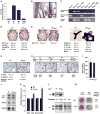
Figure 1
Figure 1. Increased bone formation, osteoid thickness, and bone resorption in Nf1ob−/− mice
A) Nf1 mRNA is expressed at higher level in differentiated osteoblasts (d4 to d10 of culture) than in TRAP+ multinucleated osteoclasts (qPCR). B) Neurofibromin immunoreactivity (brown staining) in tibia periosteal and trabecular osteoblasts. C) Nf1 cre recombination occurred in Nf1−/− osteoblasts (ob) and bones but not in osteoclasts (oc) or other cell types where Nf1 is expressed. D and E) Increased bone volume/tissue volume (BV/TV), osteoblast surface/bone surface (ObS/BS), osteoblast number/bone perimeter (Ob.nb/BPm), and bone formation rate (BFR, μm3/μm2/year) in 3- (D) and 6-month-old (E) Nf1ob−/− mice (n = 6–9, p < 0.05). F and G) Increased osteoid surface/bone surface (OS/BS), osteoid thickness (O. Th), trabecular thickness (Tb. Th), mineralization lag time (Mlt) (F), osteoclast surface/bone surface (Oc.S/BS), osteoclast number/bone perimeter (Oc.Nb/BPm), and urinary elimination of deoxypiridinoline (Dpd/creat.) (G) in 3-month-old mice Nf1ob−/− mice (n = 6–9, p < 0.05). H and I) Number of TRAP-positive multinucleated osteoclasts (Oc. Nb, red staining) in osteoblasts (Ob)/osteoclasts (BMM) cocultures (H) and M-CSF and RANKL differentiated BMM cultures (n = 3, p < 0.05) (I). J and K) α1(I) collagen (α1(I) coll, Tnsap (j, Northern), Atf4, Osterix (Osx), Runx2, and Osteocalcin (Ocn) ([K] qPCR) expression in WT and Nf1−/− osteoblasts (n > 3, p < 0.05). L) Type 1(I) collagen content in WT, Nf1−/−, and Hyp bones and osteoblasts. M) Collagen deposition (Van Gieson), alkaline phosphatase (Alk. Phos.) activity, and level in WT and Nf1−/− osteoblasts in culture (n = 3) and serum (n = 16, p < 0.05). The data represent the mean ± the SEM.
Figure 2
Figure 2. Nf1 deficiency in osteoblasts activates ATF4 via the MAPK pathway
A) Western blots. Phosphorylation of ERK1/2, RSK2, S251-ATF4, p38, AKT, and Ras activation in osteoblasts (n = 3). B and C) MEK inhibition by U0126 decreases S251-ATF4 phosphorylation ([B] Western blots) and Ocn expression ([C] qPCR) in cultured osteoblasts (n = 3, p < 0.05). D and E) MEK inhibition by PD198306 decreases osteoid parameters (OS/BS and OV/BV) (D) and collagen content ([E] Western blots) in Nf1ob−/− bones and mice (n = 7, p < 0.05). F) PKA activity in osteoblasts (n = 4, p < 0.05). G) Western blots. Phosphorylation of S254-ATF4 in osteoblasts (n = 3). H and I) qPCR. Osteoprotegerin (Opg) expression in osteoblasts ([H] n = 3, p < 0.05) and Rankl expression in immature (d2) to mature (d8) untreated or H89-treated osteoblasts ([I] n = 3, *: Nf1−/− versus wt, #: Nf1−/− versus Nf1−/− +H89, p < 0.05). The data represent the mean ± the SEM.
Figure 3
Figure 3. In vivo evidence of ATF4 involvement in Nf1ob−/− bone phenotype
A) Transgenic and endogenous Atf4 mRNA expression in WT and α1(I) collagen-Atf4 bones. B–D) BV/TV, BFR, Ob.S/BS (B), O.Th (C), Oc.S/BS and Dpd/creat. (D) in 2-month-old α1(I) collagen-Atf4 mice (n = 4, p < 0.05). E and F) Increased Ocn and Rankl expression ([E] n = 3, Northern blot) and bone α1(I) Collagen content ([F] n = 3, Western blot) in α1(I) collagen-Atf4 bones. G and H) Bone α1(I) collagen content ([G] n = 3, Western blot), O.Th ([H] n = 4, p < 0.05) in mice lacking one copy of Atf4, of Runx2 or of Osx. The data represent the mean ± the SEM.
Figure 4
Figure 4. Low-protein diet corrects RSK2-dependent bone abnormalities in Nf1ob−/− mice
A and B) GCN2 and eIF2 phosphorylation (A) and bone α1(I) Collagen content (B) in WT and Nf1ob−/− bones under ND or LPD (n = 3, Western blots). C and D) BV/TV, BFR, Ob.S/BS, Oc.S/BS and Dpd/Creat and O.Th in WT and Nf1ob−/− bones under ND or LPD (n = 6, p < 0.05). The data represent the mean ± the SEM.
Figure 5
Figure 5. High-protein diet corrects the bone abnormalities in Atf4−/− and Rsk2−/− mice
A) Percentages of Atf4−/−, Runx2−/−, and Osx−/− pups reaching 1 month of age when mothers and pups were fed ND, HFD, or HPD (Atf4:272, Runx2: 66, Osx: 60 pups counted). B and C) Bone GCN2 and eIF2a phosphorylation ([B] n = 3), BV/TV, BFR and Ob.S/BS in 1-month-old mice for each diet (except HPD-HPD/HPD-ND, 2-month-old) ([C] n = 6, p < 0.05). D) Trabeculae Von Kossa staining in long bones of E15.5 and E18.5 embryos for each diet (n = 4-8 per group, p < 0.05). E) Type I Collagen content and mRNA expression (α1(I) Col) in 1-month-old mice for each diet. F) In situ hybridization for Bsp and Ocn expression in long bone of E15.5 or E18.5 embryos for each diet. The data represent the mean ± the SEM.
Figure 6
Figure 6. Roles of neurofibromin in the regulation of bone remodeling
In osteoblasts, neurofibromin reduces Ras, MAPK and RSK2 signaling, leading to a decrease in ATF4 transcriptional activity via reduced phosphorylation of 251Serine. This leads to a reduction in protein translation and collagen synthesis. Neurofibromin also decreases PKA activity leading to a reduction of ATF4 phosphorylation at Serine 254. As a result, Rankl expression and osteoclast differentiation are normally decreased by neurofibromin. The asterisk denotes proteins whose loss causes skeletal dysplasiae treatable by dietary manipulations. Modulation of amino acid supply can correct the defects of protein translation and collagen synthesis mediated by Nf1 or RSK2 mutations.

Comment in (VSports app下载)

References

    1. Alwan S, Tredwell SJ, Friedman JM. Is osseous dysplasia a primary feature of neurofibromatosis 1 (NF1)? Clin Genet. 2005;67:378–390. - PubMed
    1. Bashey RI, Cox R, McCann J, Jimenez SA. Changes in collagen biosynthesis, types, and mechanics of aorta in hypertensive rats. J Lab Clin Med. 1989;113:604–611. - PubMed
    1. Bi W, Huang WH, Whitworth DJ, Deng JM, Zhang Z, Behringer R, de Crombrugghe B. Haploinsufficiency of Sox9 results in defective cartilage primordia and premature skeletal mineralization. Proc Natl Acad Sci USA. 2001;98:6698–6703. - PMC - PubMed
    1. Bialek P, Kern B, Yang X, Schrock M, Sosic D, Hong N, Wu H, Yu K, Ornitz DM, Olson EN, et al. A twist code determines the onset of osteoblast differentiation. Dev Cell. 2004;6:423–435. - PubMed
    1. Dacquin R, Starbuck M, Schinke T, Karsenty G. Mouse alpha1(I)-collagen promoter is the best known promoter to drive efficient Cre recombinase expression in osteoblast. Dev Dyn. 2002;224:245–251. - PubMed

Publication types

VSports - MeSH terms