Skip to main page content
U.S. flag

An official website of the United States government

Dot gov

The . gov means it’s official. Federal government websites often end in VSports app下载. gov or . mil. Before sharing sensitive information, make sure you’re on a federal government site. .

Https

The site is secure. The https:// ensures that you are connecting to the official website and that any information you provide is encrypted and transmitted securely V体育官网. .

. 2006 Aug 22;103(34):12885-90.
doi: 10.1073/pnas.0603144103. Epub 2006 Aug 15.

Severe acute respiratory syndrome coronavirus nsp1 protein suppresses host gene expression by promoting host mRNA degradation

Affiliations

Severe acute respiratory syndrome coronavirus nsp1 protein suppresses host gene expression by promoting host mRNA degradation

Wataru Kamitani et al. Proc Natl Acad Sci U S A. .

Abstract

Severe acute respiratory syndrome (SARS) coronavirus (SCoV) causes a recently emerged human disease associated with pneumonia. The 5' end two-thirds of the single-stranded positive-sense viral genomic RNA, gene 1, encodes 16 mature proteins. Expression of nsp1, the most N-terminal gene 1 protein, prevented Sendai virus-induced endogenous IFN-beta mRNA accumulation without inhibiting dimerization of IFN regulatory factor 3, a protein that is essential for activation of the IFN-beta promoter. Furthermore, nsp1 expression promoted degradation of expressed RNA transcripts and host endogenous mRNAs, leading to a strong host protein synthesis inhibition VSports手机版. SCoV replication also promoted degradation of expressed RNA transcripts and host mRNAs, suggesting that nsp1 exerted its mRNA destabilization function in infected cells. In contrast to nsp1-induced mRNA destablization, no degradation of the 28S and 18S rRNAs occurred in either nsp1-expressing cells or SCoV-infected cells. These data suggested that, in infected cells, nsp1 promotes host mRNA degradation and thereby suppresses host gene expression, including proteins involved in host innate immune functions. SCoV nsp1-mediated promotion of host mRNA degradation may play an important role in SCoV pathogenesis. .


Conflict of interest statement

Conflict of interest statement: No conflicts declared.

"V体育官网" Figures

Fig. 1.
Fig. 1.
Subcellular localization of expressed nsp1 and nsp1 in SCoV-infected cells. (A) 293 cells were transfected with pCAGGS (lane 1) or pCAGGS-nps1 (lane 2). Total intracellular proteins were extracted at 48 h after transfection, and Western blot analysis was performed by using anti-myc antibody. (B) 293T cells were transfected with pCAGGS (a) or pCAGGS nsp1 (b). 293/ACE2 cells were mock infected (c) or infected with SCoV (d). At 48 h after transfection or 8 h p.i., subcellular localization of expressed nsp1 protein and SCoV nsp1 protein was examined by using anti-myc antibody (a and b) and anti-nsp1 antibody (c and d) as primary antibodies, respectively.
Fig. 2.
Fig. 2.
Effects of nsp1 expression on SeV-induced IFN-β mRNA accumulation. 293 cells were cotransfected with IFN-β-Luc and pCAGGS (EV), IFN-β-Luc and pCAGGS-nsp1 (nsp1), or IFN-β-Luc and pCAGGS-NSs (NSs). At 24 h after transfection, cells were infected with 100 HA units/ml of SeV (+) or mock infected (−). All samples were prepared at 16 h p.i. (A) Luc activities were measured. (B) Western blot analysis of SeV N protein (SeV N) and actin. (C) The relative abundance of IFN-β mRNAs normalized to an endogenous 18S rRNA. (D) Western blot analysis of IRF-3 monomers and dimers. A and C are results of three independent experiments.
Fig. 3.
Fig. 3.
Effect of nsp1 protein expression on the reporter gene mRNA accumulation. 293 cells were independently cotransfected with pCMV-β and pCAGGS-nsp1 (A; nsp1) or pRL-SV40 and pCAGGS-nsp1 (B; nsp1). As controls, pCAGGS (EV), pCAGGS-NSs (NSs), or pCAGGS-3a (3a) were used in the place of pCAGGS-nsp1. (Upper) At 48 h after transfection, total RNAs were extracted, and Northern blot analysis was performed by using riboprobes specific for β-gal or Luc. (Lower) The same RNA samples were separated by agarose gel electrophoresis, and 28S and 18S rRNAs were stained with ethidium bromide.
Fig. 4.
Fig. 4.
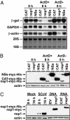
Effect of nsp1 expression on stabilities of reporter gene RNA and host endogenous mRNAs (A and B) and accumulation of nsp1 in expressing cells and SCoV-infected cells (C). (A and B) 293 cells were transfected with pCMV-β. At 16 h after transfection, cells were independently transfected with in vitro-synthesized CAT RNA transcripts (CAT), nsp1 RNA transcripts (nsp1), or NSs RNA transcripts (NSs). One hour after RNA transfection, cells were incubated with actD (ActD+) or absence of actD (ActD−). Total RNAs were extracted at 0 h (0 h) or 8 h (8 h) after actD addition. (A) Abundance of expressed β-gal RNA and endogenous GAPDH and β-actin mRNAs were examined by using Northern blot analysis. (B) Total proteins were also extracted at 0 h or 8 h after actD addition, and anti-myc antibody was used to demonstrate expression of CAT, NSs, and nsp1 proteins. (C) 293/ACE2 cells were mock-infected (Mock) or infected with SCoV (SCoV) at an moi of 3, and cell extracts were prepared at 8 h and 16 h p.i. 293 cells were transfected with pCAGGS (EV) or pCAGGS-nsp1 (nsp1), and cell extracts were prepared at 48 h after transfection (DNA). 293 cells were transfected with CAT RNA transcripts (CAT) or nsp1 RNA transcripts (nsp1), and cell extracts were prepared at 8 h after transfection (RNA). Western blot analysis was performed to detect nsp1 protein by using anti-nsp1 peptide antibody (29).
Fig. 5.
Fig. 5.
Effect of nsp1 expression of host protein synthesis. 293 cells were independently transfected with CAT RNA transcripts (CAT), nsp1 RNA transcripts (nsp1), or NSs RNA transcripts (NSs). One hour after RNA transfection, cells were incubated in the presence of actD (ActD+) or absence of actD (ActD−). Cells were labeled with 20 μCi/ml of [35S]methionine from 8.5 to 9.5 h after actD addition. Equivalent amounts of cell extracts were analyzed on a 12.5% SDS/PAGE gel. The gel was exposed to x-ray film (A) or stained with colloid Coomassie blue (B).
Fig. 6.
Fig. 6.
Effect of SCoV replication on abundance of reporter gene RNA and host endogenous mRNAs. (A) 293/ACE2 cells were transfected with pCMV-β. At 6 h after transfection, cells were mock infected (Mock) or infected with SCoV at an moi of 3 (SCoV). At 6 h and 18 h p.i., intracellular RNAs were extracted. (B) 293/ACE2 cells were transfected with pCMV-β. At 24 h after transfection, cells were mock infected (Mock) or infected with SCoV at an moi of 3 (SCoV). At 1 h p.i., intracellular RNAs were extracted (0 h) or actD was added to culture. Intracellular RNAs were extracted 14 h after actD addition (14 h). (A and B) The amounts of β-gal RNA and GAPDH and β-actin mRNAs were determined by using Northern blot analysis. Abundances of 28S and 18S rRNAs in each sample are also shown.

References

    1. Drosten C., Gunther S., Preiser W., van der Werf S., Brodt H. R., Becker S., Rabenau H., Panning M., Kolesnikova L., Fouchier R. A., et al. N. Engl. J. Med. 2003;348:1967–1976. - PubMed (V体育ios版)
    1. Ksiazek T. G., Erdman D., Goldsmith C. S., Zaki S. R., Peret T., Emery S., Tong S., Urbani C., Comer J. A., Lim W., et al. N. Engl. J. Med. 2003;348:1953–1966. - PubMed
    1. Poutanen S. M., Low D. E., Henry B., Finkelstein S., Rose D., Green K., Tellier R., Draker R., Adachi D., Ayers M., et al. N. Engl. J. Med. 2003;348:1995–2005. - PubMed
    1. Tsang K. W., Ho P. L., Ooi G. C., Yee W. K., Wang T., Chan-Yeung M., Lam W. K., Seto W. H., Yam L. Y., Cheung T. M., et al. N. Engl. J. Med. 2003;348:1977–1985. - PubMed (V体育官网)
    1. Thiel V., Ivanov K. A., Putics A., Hertzig T., Schelle B., Bayer S., Weissbrich B., Snijder E. J., Rabenau H., Doerr H. W., et al. J. Gen. Virol. 2003;84:2305–2315. - PubMed

Publication types (VSports手机版)

MeSH terms (VSports注册入口)