Skip to main page content
U.S. flag

An official website of the United States government

Dot gov

The . gov means it’s official. Federal government websites often end in . gov or . mil VSports app下载. Before sharing sensitive information, make sure you’re on a federal government site. .

Https

The site is secure. The https:// ensures that you are connecting to the official website and that any information you provide is encrypted and transmitted securely V体育官网. .

. 2006 Jul 11;103(28):10544-51.
doi: 10.1073/pnas.0603499103. Epub 2006 Jun 28.

Loss of hepatic NF-kappa B activity enhances chemical hepatocarcinogenesis through sustained c-Jun N-terminal kinase 1 activation

Affiliations

"V体育官网" Loss of hepatic NF-kappa B activity enhances chemical hepatocarcinogenesis through sustained c-Jun N-terminal kinase 1 activation

Toshiharu Sakurai et al. Proc Natl Acad Sci U S A. .

Abstract

A major link between inflammation and cancer is provided by NF-kappaB transcription factors. Ikkbeta(Deltahep) mice, which specifically lack IkappaB kinase beta (IKKbeta), an activator of NF-kappaB, in hepatocytes, are unable to activate NF-kappaB in response to proinflammatory stimuli, such as TNF-alpha. Surprisingly, Ikkbeta(Deltahep) mice are hypersusceptible to diethylnitrosamine (DEN)-induced hepatocarcinogenesis. Because defective NF-kappaB activation promotes sustained c-Jun N-terminal kinase (JNK) activation in cells exposed to TNF-alpha, whose expression is induced by DEN, and JNK activity is required for normal hepatocyte proliferation, we examined whether increased susceptibility to DEN-induced hepatocarcinogenesis in Ikkbeta(Deltahep) mice requires JNK activation. Hepatocytes express both JNK1 and JNK2, but previous studies indicate that JNK1 is more important for hepatocyte proliferation. We therefore investigated this hypothesis using mice homozygous for a JNK1 deficiency either in wild-type or Ikkbeta(Deltahep) backgrounds. In both cases, mice lacking JNK1 were much less susceptible to DEN-induced hepatocarcinogenesis. This impaired tumorigenesis correlated with decreased expression of cyclin D and vascular endothelial growth factor, diminished cell proliferation, and reduced tumor neovascularization. Whereas hepatocyte-specific deletion of IKKbeta augmented DEN-induced hepatocyte death and cytokine-driven compensatory proliferation, disruption of JNK1 abrogated this response. In addition to underscoring the importance of JNK1-mediated hepatocyte death and compensatory proliferation, these results strongly suggest that the control of tissue renewal through the IKK and JNK pathways plays a key role in liver carcinogenesis. VSports手机版.


Conflict of interest statement

Conflict of interest statement: No conflicts declared.

Figures
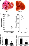
Fig. 1.
Fig. 1.
Loss of JNK1 decreases DEN-induced tumor development. (A) Livers of 10-month-old male wild-type (Wt) and Jnk1−/− mice that were given a high dose of DEN (25 mg/kg) at 15 days of age. Arrowheads, neovascularization. (B) Numbers of tumors (≥0.5 mm) and maximal tumor sizes (diameters) in livers of male wild-type (Wt, n = 10) and Jnk1−/− (n = 6) mice 10 months after DEN (25 mg/kg) injection. Horizontal bars indicate averages. (C) Numbers of tumors (≥0.5 mm) and maximal tumor sizes (diameters) in livers of male wild-type (Wt, n = 16) and Jnk1−/− (n = 15) mice 8 months after injection of a low dose of DEN (5 mg/kg). Results are means ± SE. ∗, P < 0.05 vs. wild-type mice.
Fig. 2.
Fig. 2.
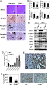
Cyclin D, VEGF, cell proliferation, and neoangiogenesis are reduced in Jnk1−/− tumors. (A) Immunohistological analysis of wild-type and Jnk1−/− HCCs. N, noncancerous liver tissues; T, tumors. Original magnification: hematoxylin/eosin (H&E), ×50; others, ×200. (B) Frequencies of proliferating (PCNA-positive), apoptotic (TUNEL-positive), and phospho-c-Jun- or phospho-ATF2-positive cells in wild-type (Wt, n = 10) and Jnk1−/− (n = 10) HCCs. Results are means ± SE. ∗, P < 0.05 vs. wild-type mice. (C) Expression of cell cycle proteins, p53, and VEGF. Lysates of microdissected HCCs (HCC; two separate samples) or nontumor liver tissue (Liver) from DEN-treated mice were gel-separated and immunoblotted with antibodies to the indicated proteins. (D) Effects of JNK1 on cyclin gene expression. RNA from tumors (T) and nontumor liver tissues (NT) were analyzed by real-time quantitative PCR, and the tumor (T)/nontumor tissue (NT) ratio of expression of the different cyclin genes was determined. Results are means ± SE (n = 4). ∗, P < 0.05 vs. wild-type mice (Wt). (E) Expression of VEGF in HCCs. Cryosections were immunostained with polyclonal VEGF antibody. N, noncancerous liver tissues; T, tumors. Original magnification: ×200. Distinction between tumor and noncancerous liver tissue was made by hematoxylin/eosin staining. (F) Intratumoral microvessel density in wild-type (Wt, n = 3) and Jnk1−/− (n = 3) HCCs. Cryosections were immunostained with anti-CD31 antibody, and microvessels were counted per high-power fields (HPF; original magnification: ×400). Data are presented as means ± SE. ∗, P < 0.05 vs. wild-type mice. Corresponding representative photomicrographs (original magnification: ×200) are shown.
Fig. 3.
Fig. 3.
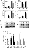
Loss of JNK1 attenuates both cell death and compensatory hepatocyte proliferation after DEN treatment. (A) Effects of JNK1 on cell death and proliferation. Mice were injected with DEN at t = 0, and alanine transaminase (ALT) levels in serum were determined at the indicated time points. The extent of liver cell apoptosis and proliferation was determined by TUNEL staining or BrdU labeling, respectively. The extent of necrosis was determined as described (3). Results are means ± SE. ∗, P < 0.05 vs. wild-type mice (Wt). (B) JNK1 contributes to DEN-induced JNK activity. Mice were treated as above, and their livers isolated when indicated and homogenized. JNK activity was determined by solid-state kinase assay with c-Jun as the substrate. Protein recovery was determined by immunoblotting with anti-JNK1 and -JNK1/2 antibodies. Ratio, relative JNK activity. (C) AP-1 DNA-binding activity in nuclear extracts of DEN-treated livers. Livers were isolated when indicated, nuclear extracts were prepared, and AP-1 activity was assessed by a mobility-shift assay. Ratio, relative AP-1 activity. (D) Effects of JNK1 on cytokine gene expression. Mice were treated as above, and liver RNA was extracted at the indicated times. Levels of cytokine mRNAs were determined by real-time quantitative PCR. Results are means ± SE (n = 4). ∗, P < 0.05 vs. wild-type mice (Wt).
Fig. 4.
Fig. 4.
JNK1 potentiates DEN-induced apoptotic cell death. (A) Effects of JNK1 on apoptotic signaling. Mice were injected with DEN, and their livers were isolated at the indicated times and homogenized. Protein extracts were gel-separated and immunoblotted with the indicated antibodies. (B) Effects of JNK1 on p53 target gene expression. Mice were treated as above, and total liver RNA was extracted at 24 h after DEN injection. Expression of p53 target genes was measured by real-time quantitative PCR. Results are means ± SE (n = 4). ∗, P < 0.05 vs. wild-type mice (Wt).
Fig. 5.
Fig. 5.
JNK1 disruption reverses increased hepatocyte death and susceptibility to HCC formation caused by the absence of hepatocyte IKKβ. (A) Numbers of tumors (≥0.5 mm) and maximal tumor sizes (diameters) in livers of male IkkβΔhep/Jnk1+/+ (Δhep, n = 10) and IkkβΔhep/Jnk1−/− (Δhep/JNK1−/−, n = 12) mice 8 months after DEN (5 mg/kg) injection. (B) Expression of cell-cycle-related proteins. Lysates of microdissected HCCs (HCC; two separate samples) or nontumor liver tissue (Liver) from DEN-treated mice were gel-separated and analyzed by immunoblotting as above. IkkβF/F mice, F/F; IkkβΔhep mice, Δhep; IkkβΔhep/Jnk1−/− mice, Δhep/JNK1−/−. (C) Effects of JNK1 on cell death and proliferation in IkkβΔhep mice. Mice of the indicated genotypes were injected with DEN, and ALT levels in serum were determined at the indicated times. The extents of liver cell apoptosis and proliferation were determined by TUNEL staining or BrdU labeling, respectively. Results are means ± SE. ∗, P < 0.05 vs. IkkβΔhep mice.

References

    1. Thorgeirsson S. S., Grisham J. W. Nat. Genet. 2002;31:339–346. - PubMed
    1. Bosch F. X., Ribes J., Diaz M., Cleries R. Gastroenterology. 2004;127:S5–S16. - PubMed (V体育平台登录)
    1. Maeda S., Kamata H., Luo J. L., Leffert H., Karin M. Cell. 2005;121:977–990. - PubMed
    1. Hansen L. J., Tennant B. C., Seeger C., Ganem D. Mol. Cell. Biol. 1993;13:659–667. - PMC - PubMed
    1. Fausto N. Semin. Liver Dis. 1999;19:243–252. - PubMed

V体育安卓版 - Publication types

MeSH terms