Skip to main page content
U.S. flag

An official website of the United States government

Dot gov

The VSports app下载. gov means it’s official. Federal government websites often end in . gov or . mil. Before sharing sensitive information, make sure you’re on a federal government site. .

Https

The site is secure V体育官网. The https:// ensures that you are connecting to the official website and that any information you provide is encrypted and transmitted securely. .

. 2006 Jun 19;173(6):949-61.
doi: 10.1083/jcb.200511072.

The V0-ATPase mediates apical secretion of exosomes containing Hedgehog-related proteins in Caenorhabditis elegans

Affiliations

The V0-ATPase mediates apical secretion of exosomes containing Hedgehog-related proteins in Caenorhabditis elegans (V体育ios版)

Samuel Liégeois et al. J Cell Biol. .

Abstract

Polarized intracellular trafficking in epithelia is critical in development, immunity, and physiology to deliver morphogens, defensins, or ion pumps to the appropriate membrane domain. The mechanisms that control apical trafficking remain poorly defined. Using Caenorhabditis elegans, we characterize a novel apical secretion pathway involving multivesicularbodies and the release of exosomes at the apical plasma membrane. By means of two different genetic approaches, we show that the membrane-bound V0 sector of the vacuolar H+-ATPase (V-ATPase) acts in this pathway, independent of its contribution to the V-ATPase proton pump activity. Specifically, we identified mutations in the V0 "a" subunit VHA-5 that affect either the V0-specific function or the V0+V1 function of the V-ATPase. These mutations allowed us to establish that the V0 sector mediates secretion of Hedgehog-related proteins VSports手机版. Our data raise the possibility that the V0 sector mediates exosome and morphogen release in mammals. .


Figures

Figure 1.
Figure 1.
VHA-5 is at the apical membrane in the excretory canal and the epidermis. (A) Drawing of the V-ATPase complex and C. elegans subunits analyzed in this study. (B) PCR analysis of wild-type, vha-5(mc38)/+, and vha-5(mc38) animals with primers in vha-5 showing that mc38 is a small deletion. (C) Western blot with a VHA-5 antiserum of wild-type C. elegans extracts (lane a) and vha-5(mc38) mutants rescued by a vha-5∷gfp transgene (lane b). A 105-kD band is visible in wild-type animals, a 135-kD band in rescued vha-5(mc38) mutants. (D) Drawing of a section through the body (left) and the epidermis (right) showing the positions of the images displayed in this and other figures. (E) Distribution of vha-5∷rfp and vha-8∷yfp in rescued vha-5(mc38) animals; XY confocal section, apical epidermal surface where the pattern appears as dots (arrowheads; excretory canal, arrows). The V1 E subunit VHA-8 (see A) colocalizes with VHA-5. (F) Immunofluorescence image of a wild-type adult with VHA-5 antiserum; VHA-5 forms dots in the epidermis. (G) Immunogold labeling against VHA-5 (gold beads, arrowheads); VHA-5 localizes mainly to apical membrane stacks of the epidermis (see also Fig. S5 D). Fig. S5 is available at http://www.jcb.org/cgi/content/full/jcb.200511072/DC1.
Figure 2.
Figure 2.
The V0 sector is specifically required for cuticle secretion. (A) Alae (arrowheads) of wild-type (WT), vha-5(mc38), and vha-4(RNAi) L1 larvae visualized by SEM; vha-5(mc38) and vha-4(RNAi) larvae have essentially no alae. (B–B″) Alae (arrowheads; B), quantification of lethality and alae defects (B′) and a representative larva filled with fluid (B″) after RNAi knockdown of the V1 subunits VHA-8 and -13, or V0 subunits VHA-1, -4, and -5; images (in B and B″) correspond to DIC micrographs. Lethality, and L1 or adult (Ad) alae defects, were quantified in separate experiments. (C) Alae (arrowheads) of a wild-type adult and an animal that survived until adulthood after RNAi against vha-5. Alae are absent in the VHA-5–defective adult. (D) GFP fluorescence of a control L1 larva carrying a vha-8∷gfp transgene (WT), and after RNAi against vha-8 (vha-8(RNAi)). The RNAi treatment almost completely removed the fluorescence in this larva, yet it has normal alae (see magnified view of the boxed area in the bottom DIC image).
Figure 3.
Figure 3.
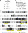
Mutations introduced in the V0 subunit VHA-5. (A) Strategy to generate vha-5 mutations based on complementation of the vha-5–null allele vha-5(mc38). (B) Predicted topology for VHA-5 based on yeast Vph1p (Nishi and Forgac, 2002) and positions of substitutions. Box, symbols for the most important phenotypes (see also Fig. S2). DbClustal alignment (http://bips.u-strasbg.fr/PipeAlign/jump_to.cgi?DbClustal+noid) of VHA-5 with the three other C. elegans “a” subunits, VHA-6 (intestinal), VHA-7 (epidermal), and UNC-32 (ubiquitous in the embryo, and then muscular and neuronal), the most closely related human and fly “a” subunits (human ATP6V0a1 and D. melanogaster V100), and the S. cerevisiae “a” subunit Vph1p. The positions of the mutations and the predicted positions of the transmembrane domains (numbered with roman letters) are indicated above VHA-5. Fig. S2 is available at http://www.jcb.org/cgi/content/full/jcb.200511072/DC1.
Figure 4.
Figure 4.
Genetic separation of the V0-specific and V0+V1 functions of the V0 subunit VHA-5. (A) Body length of adults at the same age (Error bars represent the SD; ***, significantly different from wild-type with P < 0.0001). In this and subsequent figures, vha-5(mc38) animals with a mutant vha-5∷gfp or vha-5∷rfp transgenic are noted mc38; Ex[substitution]. (B) Western blot analysis with VHA-5 antiserum of extracts prepared from the three main mutants described in the text. The VHA-5∷GFP protein levels are comparable, relative to an actin loading control. (C) Adult outer cuticle, alae (arrows), and annuli (arrowheads) observed by SEM (genotypes indicated above images). Note the stunted alae and annuli defects induced by L786S and E830Q mutations. (D) GFP fluorescence of the VHA-5 construct in the excretory canal of similar adults (top row, single XY confocal section; bottom row, transverse XZ projection. Compare the normal lumen (arrow) in the control animal with the whorls (arrowheads) induced by the W190A mutation. (E) Excretory canal in similar adults observed by TEM, and quantification of the canal section area. Note the multiple lumens (black arrowheads) in mc38; Ex[W190A] animals (dotted lines outline the excretory canal). NS, not significantly different from control animals; SD, standard deviation.
Figure 5.
Figure 5.
Knockdown of V0 and V1 subunits induces whorls in the excretory canal. VHA-5∷GFP fluorescence in the excretory canal of adults after RNAi against V0 (vha-1 and vha-4) or V1 (vha-8 and vha-13) subunits performed during larval development. Note the presence of whorls (arrowheads; the normal lumen is outlined with arrows), as in mc38; Ex[W190A] animals (Fig. 4 D).
Figure 6.
Figure 6.
Cuticle mutations impair MVB-driven exosome release. TEM micrographs of the adult epidermis. (A) Dense MVBs at low magnification (genotypes indicated above images); the number and size of MVBs are quantified below images. Note that L786S and E830Q mutations increase MVB size and number. (B) Light MVBs at higher magnification in control mc38; Ex[+] (B1–B3) and wild-type (B4) adults. Note MVB organelles (thick arrows) with intralumenal vesicles (arrowheads), in direct apposition to membrane stacks (thin arrows) at the apical epidermal plasma membrane (B1), or in apparent fusion with the plasma membrane (B2). The presence of vesicles externally (B3 and B4) suggests that these MVBs are secretory and that intralumenal vesicles become exosomes. (C) MVBs and lysosomes in mc38; Ex[E830Q] (C1–C3) and wild-type (C4) adults. MVBs are electron dense (C1), yet can evolve into normal lysosomes (C2 and C3, compare to C4). Bars, 0.5 μm. (D) Quantification of endocytic organelles in vha-5 mutants (error bars represent the SD; ***, significantly different from wild type; P < 0.0001). Cuticle mutants specifically accumulate dense and hybrid MVBs.
Figure 7.
Figure 7.
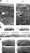
Fluid-phase endocytosis mutations do not affect cuticle formation. (A) Epidermis of a cup-5(ar465) adult visualized by TEM; note the enlarged electron-dense MVB (demarcated by dotted lines). (B) Adult alae visualized by TEM (genotypes are indicated on the left). (C) Alae of L1 larvae grown at 25°C and visualized by DIC; vps-27(ok579) is an L2 lethal mutation and rme-8(b1023) is a temperature-sensitive lethal mutation. vps-27, rme-8, and cup-5 mutations did not affect alae formation, in contrast to vha-5 cuticle mutations.
Figure 8.
Figure 8.
The V0 sector is required for the secretion of Hedgehog-related peptides through MVBs. (A) Structure of the wrt-2∷gfp construct. GFP was inserted in frame in a nonconserved region after the signal peptide cleavage site, rather than at the COOH terminus because the related WRT-1 protein undergoes autoprocessing in vitro, like Hedgehog (Porter et al., 1996). (B) XZ projections of serial confocal sections through the epidermis of adults expressing mutant VHA-5∷mRFP and WRT-2∷GFP constructs (white arrowheads, VHA-5 and WRT-2 in the epidermis; white arrows, excretory canal; part of the green signal was caused by autofluorescence, arrow on the right). The L786S mutation induced coretention of VHA-5∷mRFP and WRT-2∷GFP in homozygous (third set of images), but not in heterozygous (first set of images), vha-5(mc38) animals. The mixed mutation W327A, and the cuticle mutations L786S and V844F resulted in similar coretention phenotypes (Fig. S3 D and not depicted). (C) XZ projections of serial confocal sections through the epidermis of mc38; Ex[E830Q] adults expressing the MVB marker VPS-27∷GFP construct. (D and E) Immunogold localization of VHA-5 (D and E) and WRT-2∷GFP (D) in MVBs of mc38; Ex[E830Q] mutant (two different samples; D) and wild-type adults (E). Fig. S3 is available at http://www.jcb.org/cgi/content/full/jcb.200511072/DC1.
Figure 9.
Figure 9.
Model for apical secretion mediated by the V0 sector of the V-ATPase. We propose that in a wild-type animal (left) the V0 sector mediates fusion between the limiting membrane of MVBs and the apical plasma membrane (curved double-headed arrow). This model is supported by our genetic data, by the presence of VHA-5 at the apical plasma membrane (Fig. 1, E–G) and the MVB-limiting membrane (Fig. 8 E and Fig. S5 A), and by the accumulation of dense or hybrid MVBs in cuticle mutants (Fig. 8, Fig. S3 D, and Fig. S5, B–D). We expect the existence of two distinct V0 populations, some mediating secretion, others mediating proton pumping with the V1 sector. In cuticle defective vha-5 mutants (right), most fusion events between MVBs and the plasma membrane are compromised so that MVBs grow and become denser by accumulating their content, which can nevertheless be normally degraded.

References

    1. Amara, S.G., and M.J. Kuhar. 1993. Neurotransmitter transporters: recent progress. Annu. Rev. Neurosci. 16:73–93. - PubMed
    1. Ang, A.L., T. Taguchi, S. Francis, H. Folsch, L.J. Murrells, M. Pypaert, G. Warren, and I. Mellman. 2004. Recycling endosomes can serve as intermediates during transport from the Golgi to the plasma membrane of MDCK cells. J. Cell Biol. 167:531–543. - PMC - PubMed
    1. Aspock, G., H. Kagoshima, G. Niklaus, and T.R. Burglin. 1999. Caenorhabditis elegans has scores of hedgehog-related genes: sequence and expression analysis. Genome Res. 9:909–923. - VSports在线直播 - PubMed
    1. Bayer, M.J., C. Reese, S. Buhler, C. Peters, and A. Mayer. 2003. Vacuole membrane fusion: V0 functions after trans-SNARE pairing and is coupled to the Ca2+-releasing channel. J. Cell Biol. 162:211–222. - PMC - PubMed
    1. Beronja, S., P. Laprise, O. Papoulas, M. Pellikka, J. Sisson, and U. Tepass. 2005. Essential function of Drosophila Sec6 in apical exocytosis of epithelial photoreceptor cells. J. Cell Biol. 169:635–646. - "VSports" PMC - PubMed

Publication types

MeSH terms

Substances