Skip to main page content
U.S. flag

An official website of the United States government

Dot gov

The . gov means it’s official VSports app下载. Federal government websites often end in . gov or . mil. Before sharing sensitive information, make sure you’re on a federal government site. .

Https

The site is secure. The https:// ensures that you are connecting to the official website and that any information you provide is encrypted and transmitted securely V体育官网. .

. 2006 Jun 27;103(26):10029-34.
doi: 10.1073/pnas.0600304103. Epub 2006 Jun 16.

Rab3 GTPase-activating protein regulates synaptic transmission and plasticity through the inactivation of Rab3

Affiliations

Rab3 GTPase-activating protein regulates synaptic transmission and plasticity through the inactivation of Rab3

Ayuko Sakane et al. Proc Natl Acad Sci U S A. .

Abstract

Rab3A small G protein is a member of the Rab family and is most abundant in the brain, where it is localized on synaptic vesicles. Evidence is accumulating that Rab3A plays a key role in neurotransmitter release and synaptic plasticity. Rab3A cycles between the GDP-bound inactive and GTP-bound active forms, and this change in activity is associated with the trafficking cycle of synaptic vesicles at nerve terminals. Rab3 GTPase-activating protein (GAP) stimulates the GTPase activity of Rab3A and is expected to determine the timing of the dissociation of Rab3A from synaptic vesicles, which may be coupled with synaptic vesicle exocytosis VSports手机版. Rab3 GAP consists of two subunits: the catalytic subunit p130 and the noncatalytic subunit p150. Recently, mutations in p130 were found to cause Warburg Micro syndrome with severe mental retardation. Here, we generated p130-deficient mice and found that the GTP-bound form of Rab3A accumulated in the brain. Loss of p130 in mice resulted in inhibition of Ca(2+)-dependent glutamate release from cerebrocortical synaptosomes and altered short-term plasticity in the hippocampal CA1 region. Thus, Rab3 GAP regulates synaptic transmission and plasticity by limiting the amount of the GTP-bound form of Rab3A. .


Conflict of interest statement

Conflict of interest statement: No conflicts declared.

Figures

Fig. 1.
Fig. 1.
Targeted disruption of the Rab3 GAP p130 gene. (A) The structure of the mouse Rab3 GAP p130 gene with the first, second, and third coding exons is shown. A targeting vector was designed to remove the 3′ half of exon 1, exon 2, and the 5′ portion of the downstream intron. The construct contained 5.8 kb of the 5′ flanking sequence and 2.4 kb of the 3′ flanking sequence. The diphtheria toxin DT-A cassette was inserted at the 3′ end of the construct. In the targeted allele, the MC1-neo cassette replaces 1.2 kb of the genomic DNA. Homologous recombination in ES cells was verified by using informative restriction fragments and diagnostic probes as indicated. (B) Southern blot hybridization using the digested DNA extracted from ES cells (Upper) or mouse tails (Lower) and the 3′ external or the 5′ internal probe shown in A. The 3′ probe hybridized to the 8.5-kb wild-type or the 4.7-kb mutant fragments digested with HindIII, and the 5′ probe hybridized to the 9.7-kb wild-type or the 6.0-kb mutant fragments digested with SacI, respectively. (C) Genotyping 21-day-old mice by PCR analysis. PCR primers were selected to generate a 260-bp product indicative of the wild-type allele or a 224-bp product resulting from the disrupted p130 allele.
Fig. 2.
Fig. 2.
Biochemical and morphological analyses of the p130-deficient mice. (A) Expression of synaptic proteins in the wild-type (WT) and p130-deficient (KO) mice was analyzed by Western blotting. (B) Normal synaptic architecture in the p130-deficient brains. (Upper) Sagittal sections of hippocampi and cortices stained with hematoxylin and eosin. Arrowheads indicate the corpus callosum. (Lower) Confocal immunofluorescence analysis of frozen sections of the hippocampal CA1 and CA3 regions. Each section was double-labeled with a polyclonal antibody against synapsin I (green) and a monoclonal antibody against Rab3A (red). SO, stratum oriens; SP, stratum pyramidale; SR, stratum radiatum; SL, stratum lucidum. (C) Pull-down assays. (Left) BHK cells were transfected with Myc-tagged Rab3AQ81L or Rab3AT36N. After 48 h, the mutants were isolated by using a GST-Rim1αN-affinity column and visualized by Western blotting using an anti-Myc antibody. (Right) Wild-type or p130-deficient mouse brains were homogenized. GTP-Rab3A was then purified from the homogenates with a GST-Rim1αN-affinity column and visualized by Western blotting using an anti-Rab3A antibody.
Fig. 3.
Fig. 3.
Glutamate release from the p130-deficient synaptosomes. Glutamate release from the wild-type (WT) or p130-deficient (KO) synaptosomes by KCl-induced depolarization was measured spectrofluorometrically after the conversion of glutamate by glutamate dehydrogenase. Incubations were performed in the presence of 1.3 mM CaCl2 (Ca2+) or 0.5 mM EGTA.
Fig. 4.
Fig. 4.
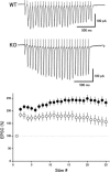
Altered activity-dependent plasticity during repetitive stimulation in the p130-deficient mice. (Upper) EPSCs in response to 25 stimuli at 14 Hz were recorded from CA1 neurons in hippocampal slices in the presence of 100 μM picrotoxin. (Lower) Relative amplitudes of EPSCs plotted as a function of stimulus number in the wild-type mice (open circles, n = 33) and mutant mice (filled circles, n = 50).
Fig. 5.
Fig. 5.
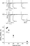
Enhanced paired-pulse facilitation in the p130-deficient mice. (Upper) EPSCs recorded from CA1 neurons in response to paired stimuli with different interstimulus intervals (50–500 ms). (Lower) Ratios of the second to the first EPSC amplitude in the wild-type mice (open circles, n = 54) and mutant mice (filled circles, n = 56) are plotted as a function of the interstimulus interval.
Fig. 6.
Fig. 6.
Unchanged MF–LTP in the p130-deficient mice. LTP was induced by 100-Hz stimuli for 1 s, and 25 μM d-AP5 was applied 5 min before the tetanic stimulation. There was no significant difference between the wild-type mice (open circles, n = 9) and mutant mice (filled circles, n = 8) in posttetanic potentiation or LTP.

VSports注册入口 - References

    1. Takai Y., Sasaki T., Matozaki T. Physiol. Rev. 2001;81:153–208. - PubMed
    1. Zerial M., McBride H. Nat. Rev. Mol. Cell Biol. 2001;2:107–117. - PubMed
    1. Takai Y., Sasaki T., Shirataki H., Nakanishi H. Genes Cells. 1996;1:615–632. - PubMed
    1. Geppert M., Südhof T. C. Annu. Rev. Neurosci. 1998;21:75–95. - VSports app下载 - PubMed
    1. Südhof T. C. Annu. Rev. Neurosci. 2004;27:509–547. - V体育平台登录 - PubMed

"V体育ios版" Publication types

"V体育ios版" MeSH terms