Skip to main page content
U.S. flag

An official website of the United States government

Dot gov

The . gov means it’s official. Federal government websites often end in . gov or . mil. Before sharing sensitive information, make sure you’re on a federal government site VSports app下载. .

Https

The site is secure V体育官网. The https:// ensures that you are connecting to the official website and that any information you provide is encrypted and transmitted securely. .

. 2006 Jul;88(1):44-51.
doi: 10.1016/j.ygeno.2006.02.012. Epub 2006 Apr 3.

Early transposable element insertion in intron 9 of the Hsf4 gene results in autosomal recessive cataracts in lop11 and ldis1 mice (V体育2025版)

Affiliations

V体育官网入口 - Early transposable element insertion in intron 9 of the Hsf4 gene results in autosomal recessive cataracts in lop11 and ldis1 mice

Elijah Talamas et al. Genomics. 2006 Jul.

V体育2025版 - Abstract

Lens opacity 11 (lop11) is an autosomal recessive mouse cataract mutation that arose spontaneously in the RIIIS/J strain. At 3 weeks of age mice exhibit total cataracts with vacuoles. The lop11 locus was mapped to mouse chromosome 8. Analysis of the mouse genome for the lop11 critical region identified Hsf4 as a candidate gene VSports手机版. Molecular evaluation of Hsf4 revealed an early transposable element (ETn) in intron 9 inserted 61 bp upstream of the intron/exon junction. The same mutation was also identified in a previously mapped cataract mutant, ldis1. The ETn insertion altered splicing and expression of the Hsf4 gene, resulting in the truncated Hsf4 protein. In humans, mutations in HSF4 have been associated with both autosomal dominant and recessive cataracts. The lop11 mouse is an excellent resource for evaluating the role of Hsf4 in transparency of the lens. .


"VSports在线直播" Figures

Fig. 1
Fig. 1
Histological sections of P21 mouse lenses. (A) lop11/lop11 shows the presence of vacuoles throughout the lens. (B) lop11/+ shows no pathological changes.
Fig. 2
Fig. 2
Chromosomal mapping of the lop11 locus. (A) Each column represents the haplotype identified in the backcross progeny: (▪) CAST/E allele, (□) RIIIS/J allele. The number of offspring inheriting each type of chromosome is listed at the bottom of each column. (B) Linkage map of the lop11 locus; the numbers on the left represent the genetic distances in centimorgans (cM).
Fig. 3
Fig. 3
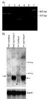
RT-PCR and Northern blot analysis. (A) Acrylamide gel electrophoresis of RT-PCR products derived from amplification of the Hsf4 gene from mouse eye mRNA. An RT-PCR product encompassing exons 1–6 is present in C57BL/6 and lop11/lop11 (lanes 1 and 2). An RT-PCR product encompassing exons 10–13 is present in C57BL/6, but absent from lop11/lop11 (lanes 4 and 5). Lanes 3 and 6 show H2O as a negative control; lane 7 is the φχ-174 RF DNA HaeIII molecular weight marker. (B) Northern analysis from P21 and P1 C57/BL6 and lop11/lop11 whole eye mRNA hybridized with a 5′ end Hsf4 probe (exons 1–6). Arrow to the left points to the wild-type Hsf4 transcript. Arrow to the right points to the lop11Hsf4 transcript of the smaller molecular weight and higher level of expression. Hybridization of the same blot with Gapdh shows even loading between samples.
Fig. 4
Fig. 4
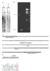
The analysis of the lop11 allele. (A) Southern blot analysis of the Hsf4 region. HindIII- and PstI-digested DNA from lop11/+ and lop11/lop11 hybridized with the 3′ end Hsf4 cDNA probe (exons 10–13). (B) LR-PCR spanning Hsf4 intron 9 showing the ETn insertion: (lane 1) lop11/lop11, (lane 2) C57BL/6, (lane 3) H2O, (lane 4) λ HindIII marker. (C) Structure of the lop11 allele. Partial genomic sequence of the chimeric Hsf4–ET genomic DNA from lop11 mice. Boxed sequences represent the primers used to amplify across the insertion in (B). Highlighted in gray is exon 10. The 5542-bp inserted sequence is the perfect match of the ETnII transposable element (Accession No. Y17106) placed in the 5′ → 3′ orientation −61 bp downstream of the 5′ intron/exon junction of exon 10. (D) Fluorescent sequence traces from the chimeric Hsf4–ET cDNA showing alternative splicing between Hsf4 exon 9 and the pseudo-exon from the ETn insertion. The dashed line separates the 3′ end of Hsf4 exon 9 and the 5′ of the pseudo-exon from the ETnII element (Accession No. Y17106).
Fig. 5
Fig. 5
Western blot analysis of protein from P21 mouse lenses from C57BL/6 and lop11/lop11. (A) Western blot using antibody specific to Hsf4; the arrow on the left points to the wild-type Hsf4 protein of about 55 kDa and the arrow on the right points to truncated Hsf4 of about 40 kDa. (B) Western blot using antibody specific for β-actin; the arrow points to the 43-kDa β-actin band.

References

    1. Foster A. Cataract—A global perspective: output, outcome and outlay. Eye. 1999;13:449–453. - PubMed
    1. Asbell PA, Dualan I, Mindel J, Brocks D, Ahmad M, Epstein S. Age-related cataract. Lancet. 2005;365:599–609. - PubMed (VSports手机版)
    1. Zetterstrom C, Lundvall A, Kugelberg M. Cataracts in children. J Cataract Refract Surg. 2005;31:824–840. - PubMed
    1. Reddy MA, Francis PJ, Berry V, Bhattacharya SS, Moore AT. Molecular genetic basis of inherited cataract and associated phenotypes. Surv Ophthalmol. 2004;49:300–315. - PubMed
    1. Francis PJ, Moore AT. Genetics of childhood cataract. Curr Opin Ophthalmol. 2004;15:10–15. - PubMed

Publication types

MeSH terms