"VSports手机版" Skip to main page content
U.S. flag

An official website of the United States government

Dot gov

The VSports app下载. gov means it’s official. Federal government websites often end in . gov or . mil. Before sharing sensitive information, make sure you’re on a federal government site. .

Https

The site is secure. The https:// ensures that you are connecting to the official website and that any information you provide is encrypted and transmitted securely. V体育官网.

Review
. 2004 Apr;85(2):47-64.
doi: 10.1111/j.0959-9673.2004.00377.x.

Smad3 as a mediator of the fibrotic response

Affiliations
Review

Smad3 as a mediator of the fibrotic response

Kathleen C Flanders. Int J Exp Pathol. 2004 Apr.

Abstract (VSports app下载)

Transforming growth factor-beta (TGF-beta) plays a central role in fibrosis, contributing to the influx and activation of inflammatory cells, the epithelial to mesenchymal transdifferentiation (EMT) of cells and the influx of fibroblasts and their subsequent elaboration of extracellular matrix. TGF-beta signals through transmembrane receptor serine/threonine kinases to activate novel signalling intermediates called Smad proteins, which modulate the transcription of target genes. The use of mice with a targeted deletion of Smad3, one of the two homologous proteins which signals from TGF-beta/activin, shows that most of the pro-fibrotic activities of TGF-beta are mediated by Smad3. Smad3 null inflammatory cells and fibroblasts do not respond to the chemotactic effects of TGF-beta and do not autoinduce TGF-beta. The loss of Smad3 also interferes with TGF-beta-mediated induction of EMT and genes for collagens, plasminogen activator inhibitor-1 and the tissue inhibitor of metalloprotease-1. Smad3 null mice are resistant to radiation-induced cutaneous fibrosis, bleomycin-induced pulmonary fibrosis, carbon tetrachloride-induced hepatic fibrosis as well as glomerular fibrosis induced by induction of type 1 diabetes with streptozotocin. In fibrotic conditions that are induced by EMT, such as proliferative vitreoretinopathy, ocular capsule injury and glomerulosclerosis resulting from unilateral ureteral obstruction, Smad3 null mice also show an abrogated fibrotic response. Animal models of scleroderma, cystic fibrosis and cirrhosis implicate involvement of Smad3 in the observed fibrosis. Additionally, inhibition of Smad3 by overexpression of the inhibitory Smad7 protein or by treatment with the small molecule, halofuginone, dramatically reduces responses in animal models of kidney, lung, liver and radiation-induced fibrosis. Small moleucule inhibitors of Smad3 may have tremendous clinical potential in the treatment of pathological fibrotic diseases VSports手机版. .


Figures
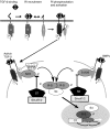
Figure 1
Figure 1
Overview of the transforming growth factor-β (TGF-β)/Smad-signalling pathway. At the cell surface, binding of TGF-β ligand to the constitutively active Type II receptor recruits the Type I receptor into the complex where it is phosphorylated. The activated Type I receptor then phosphorylates Smad 2 or 3 which are recruited there by SARA (Smad anchor for receptor activation) at the C-terminal serines. Activin also phosphorylates Smads 2/3, while BMPs phosphorylate Smads 1/5/8. The receptor-activated Smads then complex with the common mediator Smad4 and this complex translocates to the nucleus where it regulates transcription of target genes and binds to a variety of transcription factors (TFs). Activation of R-Smads by Type I receptor kinases is inhibited by Smad6 for the BMP pathway and Smad7 for the TGF-β/activin pathway. The E3 ubiquitin ligases Smurfs 1 and 2 which degrade the R-Smads also interact with Smads 6/7 and ubiquitinate the Type I receptors.
Figure 2
Figure 2
Structural organization and domain function of the three classes of Smad proteins. The overall structure of the receptor-activated and common Smad proteins comprises the conserved MH1 (grey) and MH2 (black) domains and the intervening linker (striped) region. The inhibitory Smads lack the MH1 domain. Receptor-activated Smads are phosphorylated by Type I receptor kinases on the C-terminal SSXS site. The location of proposed nuclear localization and nuclear export signals (NLS and NES, respectively) is shown. The major functions of each domain are listed on the bottom of the figure.
Figure 3
Figure 3
Proposed role of transforming growth factor-β (TGF-β)/Smad3 in wound healing and fibrosis. Degranulation of platelets releases TGF-β1 which is chemotactic for monocytes and fibroblasts with subsequent autoinduction of TGF-β by these cells and induction of collagen and other matrix proteins by fibroblasts. Processes marked with an X are blocked by loss of Smad3.
Figure 4
Figure 4
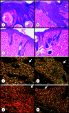
Skin of Smad3 KO mice shows less epidermal acanthosis and fibrosis than skin of WT mice 6 weeks after exposure to 30 Gy of γ-irradiation. H&E staining of irradiated WT (a and b) and KO (c and d) skin. Note the enhanced thickness of the epidermis and density of the dermis in irradiated WT compared to KO skin. Original magnification ×100. In (e–h), skin sections were stained with Picrosirius red and photographed under polarized light. The arrow marks the position of the epidermis. Non-irradiated WT (e) and KO (f) skin show similar dermal architecture. In contrast, irradiated WT (g) skin shows thicker collagen fibres with orange–red birefringence indicative of a scarring phenotype, while the architecture of the irradiated KO (h) skin more resembles that of nonirradiated skin. Original magnification ×200.
Figure 5
Figure 5
Many processes which contribute to fibrotic disease are mediated by Smad3. These include: (1) epithelial to mesenchymal transdifferentiation (EMT) which is important in the induction of renal interstitial fibrosis and proliferative vitreoretinopathy (PVR); (2) recruitment of inflammatory cells and fibroblasts and autoinduction of transforming growth factor-β (TGF-β) in these cells which is involved in induction of radiation-induced fibrosis and pulmonary fibrosis and (3) induction of collagen synthesis by TGF-β. Inhibitors of Smad3 could act at multiple sites to inhibit fibrosis.
Figure 6
Figure 6
Halofuginone treatment protects against radiation-induced leg contracture. 35 Gy irradiation was given to the right leg and leg extension compared to the nonirradiated contralateral leg of C3H/Hen female mice. Vehicle (white bars) or halofuginone (1 µg/mouse/day) (black bars) was given as a daily i.p. injection. The arrow marks the time when daily injections were stopped. *P < 0.05 compared to control.

References

    1. Aschenbrenner JK, Sollinger HW, Becker BN, Hullett DA. 1,25-(OH(2)D(3) alters the transforming growth factor beta signaling pathway in renal tissue. J. Surg. Res. 2001;100:171–175. - V体育平台登录 - PubMed
    1. Ashcroft GS, Yang X, Glick AB, et al. Mice lacking Smad3 show accelerated wound healing and an impaired local inflammatory response. Nat. Cell Biol. 1999;1:260–266. - "V体育2025版" PubMed
    1. Attisano L, Tuen Lee-Hoeflich S. The smads. Genome Biol. 2001;2:3010.1–3010.8. reviews. - "V体育平台登录" PMC - PubMed
    1. Barcellos-Hoff MH, Derynck R, Tsang ML, Weatherbee JA. Transforming growth factor-beta activation in irradiated murine mammary gland. J. Clin. Invest. 1994;93:892–899. - PMC - PubMed
    1. Border WA, Noble NA. Transforming growth factor beta in tissue fibrosis. N. Engl. J. Med. 1994;331:1286–1292. - PubMed

MeSH terms

"VSports在线直播" LinkOut - more resources