"VSports最新版本" Skip to main page content
U.S. flag

An official website of the United States government

Dot gov

The . gov means it’s official. Federal government websites often end in . gov or . mil. Before sharing sensitive information, make sure you’re on a federal government site. VSports app下载.

Https

The site is secure. The https:// ensures that you are connecting to the official website and that any information you provide is encrypted and transmitted securely V体育官网. .

Comparative Study
. 2004 Feb 15;555(Pt 1):97-114.
doi: 10.1113/jphysiol.2003.053165. Epub 2003 Oct 17.

"VSports手机版" Alternative splicing of N- and C-termini of a C. elegans ClC channel alters gating and sensitivity to external Cl- and H+

Affiliations
Comparative Study

Alternative splicing of N- and C-termini of a C. elegans ClC channel alters gating and sensitivity to external Cl- and H+

Jerod Denton (VSports app下载) et al. J Physiol. .

Abstract

CLH-3 is a meiotic cell cycle-regulated ClC Cl- channel that is functionally expressed in oocytes of the nematode Caenorhabditis elegans. CLH-3a and CLH-3b are alternatively spliced variants that have identical intramembrane regions, but which exhibit striking differences in their N- and C-termini. Structural and functional studies indicate that N- and C-terminal domains modulate ClC channel activity. We therefore postulated that alternative splicing of CLH-3 would alter channel gating and physiological functions. To begin testing this hypothesis, we characterized the biophysical properties of CLH-3a and CLH-3b expressed heterologously in HEK293 cells. CLH-3a activates more slowly and requires stronger hyperpolarization for activation than CLH-3b. Depolarizing conditioning voltages dramatically increase CLH-3a current amplitude and induce a slow inactivation process at hyperpolarized voltages, but have no significant effect on CLH-3b activity VSports手机版. CLH-3a also differs significantly in its extracellular Cl- and pH sensitivity compared to CLH-3b. Immunofluorescence microscopy demonstrated that CLH-3b is translationally expressed during all stages of oocyte development, and furthermore, the biophysical properties of the native oocyte Cl- current are indistinguishable from those of heterologously expressed CLH-3b. We conclude that CLH-3b carries the oocyte Cl- current and that the channel probably functions in nonexcitable cells to depolarize membrane potential and/or mediate net Cl- transport. The unique voltage-dependent properties of CLH-3a suggest that the channel may function in muscle cells and neurones to regulate membrane excitability. We suggest that alternative splicing of CLH-3 N- and C-termini modifies the functional properties of the channel by altering the accessibility and/or function of pore-associated ion-binding sites. .


Figures
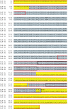
Figure 1
Figure 1. Sequence alignment of CLH-3a and CLH-3b
Regions of identity are shaded grey and non-identical regions are shaded yellow. Regions outlined in red are the presumed intracellular N- and C-termini. Cystathionine-β-synthase (CBS) domains are outlined in black. Alignment was performed using Vector NTI software (InforMax, Bethesda, MD).
Figure 2
Figure 2. Voltage-dependent properties of CLH-3a and CLH-3b
A, representative current traces from HEK293 cells cotransfected with GFP and CLH-3a or CLH-3b cDNAs. Whole-cell Cl currents were evoked by stepping membrane voltage for 1 s between −120 mV and +60 mV in 20 mV increments from a holding potential of 0 mV. Each test pulse was followed by a 1 s interval at 0 mV. B, normalized I–V relationships for CLH-3a and CLH-3b. Steady state current amplitude recorded at each test potential was normalized to that measured at −120 mV. Values are means ± s.e. (n = 11). C, Boltzmann fits of normalized I–V relationships for ICLH-3a and ICLH-3b. Fits were performed using the equation I(Vm) = ([A1A2]/[1 +e(Vm − V0.5)/k]) + A2, where V0.5 is the half-activation potential and k is the slope factor.
Figure 3
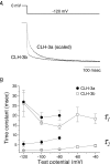
Figure 3. Kinetics of CLH-3a and CLH-3b activation
A, representative current traces showing activation kinetics of CLH-3 splice variants at −120 mV. The ICLH-3a trace was scaled by a factor of 1.7 and superimposed over the ICLH-3b trace for comparison of activation time courses. B, slow (τ1) and fast (τ2) time constants for CLH-3a and CLH-3b. Time constants were derived by fitting the first 50 ms of hyperpolarization-induced current activation with a double exponential function. Activation of CLH-3a was nominal at −60 mV (Fig. 2B) and time constants could be accurately derived only at more hyperpolarized voltages. Values are means ± s.e. (n = 8–9).
Figure 4
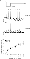
Figure 4. Time-dependent effects of predepolarization on hyperpolarization-induced activation of CLH-3a and CLH-3b
A, transfected HEK293 cells were voltage clamped for 1–7 s at 60 mV from a holding potential of 0 mV and then stepped to −120 mV for 2 s to activate the expressed channels. B, time dependency of predepolarization on peak ICLH-3a and ICLH-3b amplitude measured between 170 ms and 270 ms after the onset of the −120 mV test pulse. Currents are plotted relative to those measured from the holding potential of 0 mV (i.e. I0 mV). Values are means ± s.e. (n = 6).
Figure 5
Figure 5. Voltage-dependent effects of predepolarization on hyperpolarization-induced activation and inactivation CLH-3a and CLH-3b
A, representative current traces at −120 mV from CLH-3a- and CLH-3b-transfected HEK293 cells. Cells were voltage clamped for 3 s at condition potentials (CP) from −20 mV to 60 mV and then stepped to −120 mV for 2 s to activate the expressed channels. B, effect of conditioning potential on hyperpolarization-induced activation of ICLH-3a and ICLH-3b. Peak current amplitude was measured between 170 ms and 270 ms after stepping to −120 mV. Current values were normalized to that measured following a conditioning pulse of −20 mV (i.e. I−20 mV). Values are means ± s.e. (n = 8–17). C, effect of conditioning potential on inactivation of ICLH-3a and ICLH-3b. Mean normalized pseudo-steady-state current (ISS) was measured over the last 20 ms of the −120 mV test pulse and normalized to peak current (Ipeak) amplitude. Values are means ± s.e. (n = 8–17).
Figure 6
Figure 6. Effects of predepolarization on time constants of activation and inactivation of CLH-3a
CLH-3a-expressing HEK293 cells were voltage clamped for 3 s at condition potentials from −20 mV to 60 mV and then stepped to −120 mV for 2 s to activate the channel. Slow (τ1) and fast (τ2) activation time constants were derived by fitting the first 50 ms of hyperpolarization-induced current activation with a double exponential function. The inactivation time constant (τ) was derived by fitting the entire time course of current inactivation with a single exponential function. Values are means ± s.e. (n = 9).
Figure 7
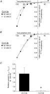
Figure 7. Effects of extracellular Cl on CLH-3a and CLH-3b
A and B, representative I–V relationships for ICLH-3a and ICLH-3b respectively, recorded in 92 mm Cl control bath solution and at 30 s after switching to a 12 mm Cl bath solution. Current values measured at each test potential were normalized to that measured at −120 mV in 92 mm Cl bath solution. C, relative amplitude for ICLH-3a and ICLH-3b measured at −100 mV in 92 mm Cl bath solution versus 12 mm Cl. Values are means ± s.e. (n = 5–7). *P < 0.004 compared to CLH-3a.
Figure 8
Figure 8. Effect of extracellular acidification on CLH-3a and CLH-3b
A and B, representative I–V relationships for ICLH-3a and ICLH-3b respectively, recorded in pH 7.4 control bath and at 30 s after switching to pH 5.9, pH 6.5, or pH 8.1 bath solutions. C, effects of changes in extracellular pH on ICLH-3a and ICLH-3b measured at −100 mV. Values are means ± s.e. (n = 4–12). D, voltage dependence of the pH 6.5-induced activation of ICLH-3a and ICLH-3b. Values are means ± s.e. (n = 8–12).
Figure 9
Figure 9. Activation kinetics of native ICLH-3
A, representative whole-cell Cl current traces under basal conditions and after maximal activation by swelling in an oocyte isolated from a wild-type (WT) worm and in an oocyte isolated from a clh-3(ok763) mutant worm. The clh-3(ok763) oocyte was swollen for 10 min. Currents were evoked by stepping membrane voltage for 1 s between −100 mV and +60 mV in 20 mV increments from a holding potential of 0 mV. Each test pulse was followed by a 1 s interval at 0 mV. The corresponding I–V relationships for these currents are shown in B. C, time constants for hyperpolarization-induced activation of native ICLH-3a in swollen oocytes. Values are means ± s.e. (n = 4–8).
Figure 10
Figure 10. Voltage- and time-dependent effects of predepolarization on hyperpolarization-induced activation of native ICLH-3
A, representative whole-cell current traces at −120 mV in a swollen wild-type oocyte. The oocyte was voltage clamped for 3 s at conditioning potentials (CP) from −20 mV to 60 mV and then stepped to −120 mV for 1 s to activate native ICLH-3. B, effect of predepolarization on native ICLH-3 at −120 mV under basal conditions and after current activation by oocyte swelling, oocyte meiotic maturation (MM) or ATP depletion. Peak current amplitudes were quantified between 170 ms and 270 ms after the onset of the −120 mV test pulse following a predepolarization of −20 mV or 60 mV. Values are means ± s.e. (n = 3–12). C, time-dependence of predepolarization on native ICLH-3 in a swollen oocyte. Oocytes were voltage clamped for 1–7 s at 60 mV from a holding potential of 0 mV and then stepped to −120 mV for 1 s to activate native ICLH-3. D, Mean ICLH-3 amplitude at −120 mV in swollen oocytes plotted as a function of the time they were clamped at a conditioning potential of 60 mV. Currents are plotted relative to those measured from the holding potential of 0 mV (i.e. I0 mV). Data are shown on the same Y-axis scale as Fig. 4B to facilitate comparison with heterologously expressed splice variants. Values are means ± s.e. (n = 3).
Figure 11
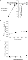
Figure 11. Effect of extracellular Cl and pH on native ICLH-3
A, I–V relationships of swelling-activated native ICLH-3 in 124 mm Cl control saline and at 30 s after switching to a 16 mm Cl bath solution. Values are means ± s.e. (n = 7). B, relative change in native ICLH-3 current amplitude after switching from a pH 7.3 bath solution to pH 5.9, pH 6.5, or pH 8.1 bath solutions. The data have been plotted on the same scale as Fig. 8C for comparison with ICLH-3a and ICLH-3b. Values are means ± s.e. (n = 4–7). C, voltage dependence of native ICLH-3 activation by bath acidification to pH 6.5. Relative current values are plotted on the same Y-axis scale used in Fig. 8D for comparison with heterologously expressed splice variants. Values are means ± s.e. (n = 7).
Figure 12
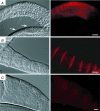
Figure 12. Immunolocalization of CLH-3b in the C. elegans gonad
Differential interference contrast (left panels) and fluorescence (right panels) micrographs of isolated C. elegans gonads. A, section of distal (arrows) and beginning of proximal gonad. CLH-3b immunostaining is first detected in the loop region of the gonad where germ cell nuclei are cellularized. B, section of proximal gonad containing single oocytes. C, section of proximal gonad isolated from a worm injected with clh-3 dsRNA. Faint background staining is similar to that observed in gonads immunoreacted with preimmune serum and in gonads in which CLH-3b antiserum was competed with CLH-3b fusion protein (data not shown). Fluorescence image exposure time is 300 ms in A, 200 ms in B and 1 s in C. Antiserum was diluted 1 : 50 (A) or 1 : 200 (B and C). Scale bars are 20 μm.

References

    1. Adrian RH, Bryant SH. On the repetitive discharge in myotonic muscle fibres. J Physiol. 1974;240:505–515. - V体育官网入口 - PMC - PubMed
    1. Arreola J, Begenisich T, Melvin JE. Conformation-dependent regulation of inward rectifier chloride channel gating by extracellular protons. J Physiol. 2002;541:103–112. - PMC - PubMed
    1. Bargmann CI. Neurobiology of the Caenorhabditis elegans genome. Science. 1998;282:2028–2033. - PubMed
    1. Brenner S. The genetics of Caenorhabditis elegans. Genetics. 1974;77:71–94. - PMC - PubMed
    1. Chen TY, Miller C. gating and voltage dependence of the ClC-0 Cl− channel. J General Physiol. 1996;108:237–250. - PMC (V体育安卓版) - PubMed

MeSH terms (V体育官网入口)

V体育平台登录 - LinkOut - more resources