Skip to main page content
U.S. flag

An official website of the United States government

Dot gov

The VSports app下载. gov means it’s official. Federal government websites often end in . gov or . mil. Before sharing sensitive information, make sure you’re on a federal government site. .

Https

The site is secure. The https:// ensures that you are connecting to the official website and that any information you provide is encrypted and transmitted securely V体育官网. .

. 2003 Nov 1;17(21):2648-63.
doi: 10.1101/gad.1144003. Epub 2003 Oct 16.

Transcriptional activation via sequential histone H2B ubiquitylation and deubiquitylation, mediated by SAGA-associated Ubp8 (VSports在线直播)

Affiliations

Transcriptional activation via sequential histone H2B ubiquitylation and deubiquitylation, mediated by SAGA-associated Ubp8

Karl W Henry et al. Genes Dev. .

"V体育2025版" Abstract

Gene activation and repression regulated by acetylation and deacetylation represent a paradigm for the function of histone modifications. We provide evidence that, in contrast, histone H2B monoubiquitylation and its deubiquitylation are both involved in gene activation. Substitution of the H2B ubiquitylation site at Lys 123 (K123) lowered transcription of certain genes regulated by the acetylation complex SAGA. Gene-associated H2B ubiquitylation was transient, increasing early during activation, and then decreasing coincident with significant RNA accumulation. We show that Ubp8, a component of the SAGA acetylation complex, is required for SAGA-mediated deubiquitylation of histone H2B in vitro. Loss of Ubp8 in vivo increased both gene-associated and overall cellular levels of ubiquitylated H2B. Deletion of Ubp8 lowered transcription of SAGA-regulated genes, and the severity of this defect was exacerbated by codeletion of the Gcn5 acetyltransferase within SAGA VSports手机版. In addition, disruption of either ubiquitylation or Ubp8-mediated deubiquitylation of H2B resulted in altered levels of gene-associated H3 Lys 4 methylation and Lys 36 methylation, which have both been linked to transcription. These results suggest that the histone H2B ubiquitylation state is dynamic during transcription, and that the sequence of histone modifications helps to control transcription. .


Figures (V体育官网)

Figure 1.
Figure 1.
Role of ubiquitylated H2B in the expression of SAGA-dependent genes. (A) GAL1 and SUC2 transcription in HTB1 and htb1-KR cells. RNA was analyzed by S1 nuclease protection assay. Fold induction was calculated as the level of expression under inducing conditions (I) compared with repressing conditions (R) and is presented as the percentage of wild-type induction (set at 100%). tRNA was used as a loading control for the gene-specific RNA. The repressing and inducing conditions were, respectively, GAL1, 2% glucose and 2% galactose for 2.5 h at OD600 nm = 0.8; SUC2, 2% glucose and 0.05% glucose for 2.5 h at OD600 nm < 0.5. The strains were wild type (HTB1+) and htb1-KR, bearing a lysine-to-arginine substitution at residue 123, the known H2B ubiquitylation site (Robzyk et al. 2000). Quantitation was done by PhosphorImager analysis. (B) Double chromatin immunoprecipitation (ChDIP) of ubH2B and RNA analysis during galactose induction. The histogram represents ChDIP analysis, and the line graph represents RNA analysis. Formaldehyde cross-linked chromatin was obtained from wild-type (left) and htb1-KR (right) strains bearing Flag-tagged H2B and HA-tagged ubiquitin in glucose medium (Glu) or during the indicated time course in galactose-containing medium. Sonicated chromatin was immunoprecipitated first with anti-Flag antibody (M2, Sigma) and eluted with 3× Flag peptide (Sigma). Eluates were then immunoprecipitated with anti-HA antibody (12CA5, Roche) before elution. Quantitative PCR analysis of eluted DNA was done in real time. The level of immunoprecipitated chromatin from the GAL1 promoter (white bars) and Int. V region (black bars) is shown as relative immunoprecipitation, a ratio of immunoprecipitated material to input chromatin (left Y-axis). GAL1 RNA levels were assayed in the same samples. RNA was isolated from wild-type (left) or htb1-KR (right) samples and analyzed by S1 analysis. GAL1 expression was normalized to tRNA levels and is presented as fold change in expression relative to expression in glucose (right Y-axis). (C) ubH2B ChDIP and GAL1 RNA analysis in wild-type cells during derepression and activation. Treatments were the same as in B except that cells were incubated in raffinose (GAL1 derepressing) for 2 h prior to the addition of galactose. Samples were taken in glucose and then at indicated times in raffinose and in galactose. Data are normalized to input and Int. V levels and are presented as fold relative IP with the glucose input-normalized immunoprecipitated values set at 1.0 and all others compared with these samples. RNA was treated as in B.
Figure 2.
Figure 2.
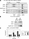
Analysis of Ubp8 association within Ada2-containing complexes. (A) Chromatographic fractionation of Ubp8 from whole-cell extracts. Cell extracts containing Ada2-TAP and Ubp8-Flag were fractionated first via the TAP purification method (Puig et al. 2001) followed by MonoQ ion exchange chromatography. Even-numbered fractions (14-42) from the MonoQ column were subjected to Western blotting to detect Ubp8-Flag, which was compared with ADA/SALSA/SAGA-associated (as indicated) Ada3 and Gcn5, SALSA/SAGA-associated Spt3, and SAGA-specific Spt8. Samples from inputs (IN) and flowthroughs (FT) for calmodulin-bead binding (C) and MonoQ column (M) purification steps were included. (B) Association of Ubp8 in ADA or SAGA complexes. ADA and SAGA fractions eluted from the MonoQ column were immunoprecipitated with anti-Flag affinity resin (Sigma), and subjected to SDS-PAGE and Western blotting to detect Ubp8-Flag, Ada3, and Gcn5. Input represents 30% of material in the immunoprecipitation. (C) ChIP analysis of Gcn5 and Ubp8 at the GAL1 promoter. Gcn5-3HA (open bars) and Ubp8-3HA (closed bars) binding in a wild-type background were analyzed by ChIP at the GAL1 promoter in glucose (0 time point) and in galactose (60 and 120 min time points). This association was compared with the Int. V region [Gcn5-3HA sample (open bars, black stripes) or Ubp8-3HA (closed bars, white stripes)]. Data are presented as fold relative IP with glucose input-normalized immunoprecipitated values set to 1.0 and all others compared with these samples.
Figure 3.
Figure 3.
H2B ubiquitylation in UBP8 and ubp8Δ strains. (A) Modified H2B in wild-type and ubp8Δ strains. Anti-Flag immunoprecipitates from N-terminally Flag-tagged H2B in wild-type and indicated mutant strains were evaluated by Western blot analysis to determine the relative levels of ubiquitin-modified and unmodified Flag-H2B as described previously (Robzyk et al. 2000). The antibodies used for Western blot analysis are listed in Table 3. Detection of ub-Flag-H2B with anti-Flag antibody and anti-ubiquitin antibody was done in separate Western blots, and their migration was comparable as indicated by molecular weight standards (shown in kilodaltons). (B) Ubiquitylation of H2B in rad6Δ, spt20Δ, and ubp3Δ strains. Western blotting was performed as in A with anti-Flag immunoprecipitated lysates from the indicated strains. The location of molecular weight standards is indicated. (C) In vitro deubiquitylation of ubH2B by Ubp8 within SAGA. SAGA was purified from UBP8 and ubp8Δ strains, and equivalent amounts (see Fig. 5B) were incubated for 30 min with ubH2B and H2B, which bore either single or double tags as indicated. ubH2B and H2B were obtained by anti-Flag immunoprecipitation from ubp8Δ cell lysates. The input was a mock-treated ubH2B sample. Western blotting was performed with anti-Flag (left; to detect H2B and ubH2B containing both tagged and untagged ubiquitin) or with anti-HA (right; to detect ubiquitin-modified H2B and free ubiquitin). (Lower panel, right) Intact HA-ubiquitin (judged from molecular weight standards) that was hydrolyzed from ubH2B by the SAGA UBP8 sample, but not from SAGA ubp8Δ. (D) ChDIP analysis of ubH2B at the GAL1 promoter in ubp8Δ. Data represent ChDIP analysis for ubH2B in both wild-type (open boxes, solid line) and ubp8Δ (closed boxes, broken line) backgrounds as performed in Figure 1C.
Figure 5.
Figure 5.
Effect of loss of Ubp8 on SAGA function and Gal4 stability. (A) SAGA stability in wild-type and ubp8Δ strains. (Left) Immunoblots of IgG-immunoprecipitated samples obtained from Ada2-TAP-tagged strains in which Ubp8 was double-Flag-tagged (WT) or deleted (ubp8Δ). Equivalent amounts of protein were used for each immunoprecipitation. (Right) SAGA was purified via standard TAP procedure followed by MonoQ ion exchange chromatography (as in Fig. 2A). Complexes from wild-type and ubp8Δ strains were analyzed by Western blotting using twofold serially diluted samples of each purified SAGA complex. (B) HAT activity of SAGA derived from wild-type or ubp8Δ strains. Threefold serial dilutions of equivalent amounts of wild-type (white bars) or ubp8Δ (gray bars) SAGA complex were assayed for HAT activity on core histones (Sigma) as described previously (John et al. 2000). Background (black bars) indicates incorporation of 3H-acetate without added SAGA complex. (C) Gal4 stability in wild-type and ubp8Δ strains. The Gal4 activator was triple HA-tagged in the wild-type or ubp8Δ backgrounds. Equivalent amounts of protein from lysates collected in glucose (0 min) or galactose-induced cultures (30, 60, and 90 min) were immunoprecipitated with anti-HA antibody and analyzed by Western blotting with an anti-Gal4 DNA-binding domain antibody (Table 3).
Figure 4.
Figure 4.
Role of Ubp8 in SAGA-dependent expression. (A) Cell growth and expression in ubp8Δ. (Upper panels) Fivefold serial dilutions of exponentially growing yeast were spotted on plates containing rich medium, with glucose (YPD), galactose (YP-galactose; GAL1-inducing), or ethanol/glycerol (YP-EtOH/Gly; ADH2-inducing). Wild-type or indicated mutant strains were plated and incubated at 30°C for 48 h. (Lower panels) RNA was extracted from cell pellets collected at the indicated times during incubation in galactose (inducing for GAL1) or ethanol/glycerol (inducing for ADH2) in the indicated strains. Expression was analyzed by RT-PCR in real time and normalized to ACT1 levels. Data are presented as fold induction with wild-type expression in glucose medium set at 1.0. (B) Genetic interaction of ubp8Δ is specific to gcn5Δ and not sas3Δ. Fivefold serial dilutions of the indicated strains were treated as in A. (C) Analysis of Ubp8 catalytic mutant. The ubp8Δ gcn5Δ mutant was transformed with plasmids that contained GCN5 (promoter and open reading frame; pGCN5) and either a wild-type UBP8 gene (pUBP8) or an allele bearing a substitution mutation in the putative catalytic site of Ubp8 (pubp8C146S). Wild type and ubp8Δ gcn5Δ were also transformed with vectors alone. Fivefold serial dilutions of exponentially growing cells were spotted onto selective plates as indicated and grown at 30°C for 48 h.
Figure 6.
Figure 6.
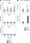
Effect of altered histone H2B ubiquitylation on histone H3 methylation status. (A) Analysis of H3 K4 methylation in htb1-KR or ubp8Δ strains. ChIP analysis of the GAL1 promoter in wild-type and ubp8Δ strains. Antibodies specific for different methylation states of histone H3 K4 (3Me, top; 2Me, middle; and 1Me, bottom). Samples were taken from wild-type (open bars), ubp8Δ (gray bars), and htb1-KR (black bars) strains grown in glucose (Glu) and at the times indicated after transfer to galactose medium. The data represent the amplification product from immunoprecipitated material normalized to input material and compared with wild-type glucose IP levels (set as 1.0). (B) Histone H3 methylation of K36. ChIP analysis of the strains used in A was performed using antibodies to H3 K36 dimethylated as in A. The upper panel shows wild type and ubp8Δ; the lower panel shows wild type and htb1-KR. Cells were grown in glucose-containing medium (Glu), washed, and transferred to raffinose medium for 2 h prior to the addition of galactose (Gal).
Figure 7.
Figure 7.
Model of the role of ubiquitylation and deubiquitylation at GAL1. Schematic representation of the GAL1 promoter and a nucleosome over the TATA/5′ end of the ORF under poised (A; Ub + K4Me) and activated (B; K4Me + K36Me) conditions, showing the relationship between ubiquitylation/deubiquitylation and methylation in the wild-type strain. Paf1 complex loads both Set1 (K4Me) and Set2 (K36Me). The lower cartoons represent altered ubiquitylation and methylation in ubp8Δ and htb1-KR backgrounds. (C) In the absence of H2B ubiquitylation, there is no K4 methylation and high K36 methylation. (D) In the absence of Ubp8, there is high K4 methylation and lowK36 methylation. These altered ubiquitylation/methylation patterns lead to lowtranscription.

References

    1. Amerik A.Y., Li, S.-J., and Hochstrasser, M. 2000. Analysis of the deubiquitinating enzymes of the yeast Saccharomyces cerevisiae. Biol. Chem. 381: 981-992. - PubMed
    1. Belotserkovskaya R., Sterner, D.E., Deng, M., Sayre, M.H., Lieberman, P.M., and Berger, S.L. 2000. Inhibition of TATA-binding protein function by SAGA subunits Spt3 and Spt8 at Gcn4-activated promoters. Mol. Cell. Biol. 20: 634-647. - PMC - PubMed
    1. Bernstein B.E., Humphrey, E.L., Erlich, R.L., Schneider, R., Bouman, P., Liu, J.S., Kouzarides, T., and Schreiber, S.L. 2002. Methylation of histone H3 Lys 4 in coding regions of active genes. Proc. Natl. Acad. Sci. 99: 8695-8700. - PMC - PubMed
    1. Bhaumik S.R. and Green, M.R. 2001. SAGA is an essential in vivo target of the yeast acidic activator Gal4p. Genes & Dev. 15: 1935-1945. - "VSports手机版" PMC - PubMed
    1. Boeger H., Griesenbeck, J., Strattan, J.S., and Kornberg, R.D. 2003. Nucleosomes unfold completely at a transcriptionally active promoter. Mol. Cell 11: 1587-1598. - PubMed

Publication types