Skip to main page content
U.S. flag

An official website of the United States government

Dot gov

The VSports app下载. gov means it’s official. Federal government websites often end in . gov or . mil. Before sharing sensitive information, make sure you’re on a federal government site. .

Https

The site is secure V体育官网. The https:// ensures that you are connecting to the official website and that any information you provide is encrypted and transmitted securely. .

. 2003 Nov;9(11):1371-82.
doi: 10.1261/rna.5520403.

The Saccharomyces cerevisiae U2 snRNA:pseudouridine-synthase Pus7p is a novel multisite-multisubstrate RNA:Psi-synthase also acting on tRNAs (V体育ios版)

Affiliations

The Saccharomyces cerevisiae U2 snRNA:pseudouridine-synthase Pus7p is a novel multisite-multisubstrate RNA:Psi-synthase also acting on tRNAs

Isabelle Behm-Ansmant et al. RNA. 2003 Nov.

Abstract

The Saccharomyces cerevisiae Pus7 protein was recently characterized as a novel RNA:pseudouridine (Psi)-synthase acting at position 35 in U2 snRNA. However, U2 snRNA was the only potential substrate tested for this enzyme. In this work, we demonstrated that although Pus7p is responsible for the formation of only one of the six Psi residues present in yeast UsnRNAs, it catalyzes U to Psi conversion at position 13 in cytoplasmic tRNAs and at position 35 in pre-tRNA(Tyr). Sites of RNA modification by Pus7p were identified by analysis of the in vivo RNA modification defects resulting from the absence of active Pus7p production and by in vitro tests using extracts from WT and genetically modified yeast cells. For demonstration of the direct implication of Pus7p in RNA modification, the activity of the WT and mutated Pus7p recombinant proteins was tested on in vitro produced tRNA and pre-tRNA transcripts VSports手机版. Mutation of an aspartic acid residue (D256) that is conserved in all Pus7 homologs abolishes the enzymatic activity both in vivo and in vitro. This suggests the direct involvement of D256 in catalysis. Target sites of Pus7p in RNAs share a common sequence Pu(G/C)UNPsiAPu (Pu = purine, N = any nucleotide), which is expected to be important for substrate recognition. Modification of tRNAs by Pus7p explains the presence of Pus7p homologs in archaea and some bacteria species, which do not have U2 snRNA, and in vertebrates, where Psi34 (equivalent to Psi35 in yeast) formation in U2 snRNA is an H/ACA snoRNA guided process. Our results increase the number of known RNA modification enzymes acting on different types of cellular RNAs. .


"VSports" Figures
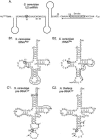
FIGURE 1.
FIGURE 1.
Sequences and secondary structures of the Pus7p RNA substrates studied in this work. (A) The 5′-terminal part of S. cerevisiae U2 snRNA containing the three identified Ψ residues and the Sm site (Massenet et al. 1999). (B) The S. cerevisiae tRNAAsp (B1) and tRNAGlu (B2). (C) The S. cerevisiae (C1) and A. thaliana (C2) pre-tRNATyr are drawn with all their identified post-transcriptional modifications (tRNA database; Sprinzl et al. 1998; see also Internet site http://www.uni-bayreuth.de/departments/biochemie/sprinzl/trna/). The intronic sequences in pre-tRNAs are shown in small characters and arrows indicate exon–intron borders. The Ψ residues found to be formed by Pus7p are circled. Oligonucleotides used for primer extension analysis are indicated. In the yeast pre-tRNATyr transcript the C1–G72 base pair (boxed) was converted into a G1–C72 base pair to increase transcription efficiency.
FIGURE 2.
FIGURE 2.
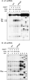
Disruption of the PUS7 gene does not alter formation of Ψ residue in U1 (A) and U5 snRNAs (B). Total RNA was extracted from the WT and mat-a ΔPUS7 S. cerevisiae BY4742 strains and modified by CMCT, for 1, 10, and 20 min with (+) or without (−) subsequent alkaline treatment (OH). A control experiment was performed in the absence of CMCT treatment. Lanes U, G, C, and A correspond to the sequencing ladders obtained with the same oligonucleotide. The reverse transcription stops, corresponding to residues Ψ5 and Ψ6 (A) and to Ψ99 (B) are indicated by arrows.
FIGURE 3.
FIGURE 3.
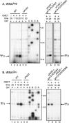
Ψ13 formation in cytoplasmic tRNAAsp (A) and tRNAGlu (B) is abolished upon PUS7 gene disruption and restored by complementation with plasmid p413GalS-PUS7. Total RNA was extracted from the WT and mat-a ΔPUS7 strains and modified by CMCT, for 1, 10, and 20 min with (+) or without (−) subsequent alkaline treatment (OH). A control experiment was performed in the absence of CMCT treatment. Lanes U, G, C, and A correspond to the sequencing ladders obtained with the same oligonucleotide. The reverse transcription stops, corresponding to residues Ψ13, are indicated by arrows. To verify the direct involvement of Pus7p in the U to Ψ conversion at position 13 of tRNAs, the ΔPUS7 strain was transformed with plasmid p413GalS-PUS7, bearing the wild-type (WT) or mutated (D256A) PUS7 gene. Total RNA was extracted from these two transformed strains, and tRNAAsp (A) and tRNAGlu (B) were analyzed by the CMCT/RT approach (shown in lanes 7, 8, 9, and 10 in A and B).
FIGURE 4.
FIGURE 4.
Tests of the tRNA:Ψ1, Ψ13 and Ψ35-synthase activities in different yeast S10 extracts. In vitro transcribed RNA substrates labeled by incorporation of [α-32P]ATP (yeast tRNAAsp, A, yeast pre-tRNATyr, B) or [α-32P]UTP (yeast tRNAArg, C) were incubated with different S10 extracts in the conditions described in Materials and Methods. Extracts were prepared from cells of the WT BY4742 strain (WT), the isogenic ΔPUS7 strain, and the ΔPUS7 strain complemented with the WT or D256A variant PUS7 gene. The activity on yeast pre-tRNATyr of an extract from the ΔPUS1 BY4742 strain was also tested. The tRNAAsp and tRNAArg transcripts incubated in the same conditions but in the absence of S10 extract, were used as controls. After incubation, the transcripts were digested with T2 RNase and 3′NMPs were fractionated on TLC as described in Materials and Methods. The autoradiograms of the TLC plates are shown. Positions of the NMPs (Ap, Cp, Up, Gp) and ΨMP nucleotides were identified according to Keith (1995). Quantification of the Ψ residue formation was done by measuring the radioactivity in each spot with a PhosphoImager and the ImageQuant software.
FIGURE 5.
FIGURE 5.
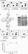
Recombinant His6–Pus7p catalyzes specifically U to Ψ conversion at position 13 in yeast cytoplasmic tRNAAsp. (A) CMCT/RT mapping of Ψ residues formed upon incubation of tRNAAsp with His6–Pus7p. Cold in vitro produced tRNAAsp was incubated in the presence (+) or absence (−) of His6–Pus7p in the conditions described in Materials and Methods. The modified tRNAAsp was analyzed by the CMCT/RT approach (same legend as in Fig. 3 ▶). (B) Analysis of Ψ13 formation in tRNAAsp by the nearest neighbor approach. tRNAAsp transcript labeled with [α-32P]ATP was incubated with the recombinant His6–Pus7p or the His6–Pus7D256Ap mutant in the conditions described in Materials and Methods. A control incubation was performed in the absence of the recombinant protein. The incubated tRNAsAsp were digested with T2 RNase and the released 3′NMPs were fractionated by TLC, as described in Materials and Methods. The autoradiograms of the TLC plates are shown. The molar ratio of Ψ residues formed in tRNAs, as deduced from quantification of the radioactivity of the spots, is given at the bottom of the panels. (C) Same experiment as in (B) with the U13C variant tRNAAsp. (D) Time-course analysis of tRNAAsp modification by the recombinant His6–Pus7p enzyme. tRNAAsp transcript (50–100 fmoles) was incubated with 750 fmoles of His6–Pus7p in the conditions described in Materials and Methods. Aliquot fractions were collected at the indicated times after the beginning of the incubation. For each fraction, RNA was digested with T2 RNase and the released products were analyzed by 2D TLC. Confidence intervals are calculated taking into account the relative radioactivity measured for the ΨMP spot and the Ap, Cp, Gp, and Up spots on 2D TLC.
FIGURE 6.
FIGURE 6.
Recombinant His6–Pus7p catalyzes specifically the U to Ψ conversion at position 35 in yeast pre-tRNATyr. (A) Analysis of Ψ35 formation in pre-tRNATyr by the nearest neighbor approach. Yeast pre-tRNATyr uniformly labeled with [α-32P]ATP was incubated with the recombinant His6–Pus7p or His6–Pus7D256Ap variant in the conditions described in Materials and Methods. A control experiment was performed in the absence of recombinant protein. Modified and unmodified pre-tRNAsTyr were digested with T2 RNase and the 3′NMPs were fractionated by TLC. The autoradiograms of the TLC plates are shown. The molar ratio of Ψ residues formed in tRNAs, as deduced from quantification of the radioactivity of the spots, is given at the bottom of the panels. (B) Mapping of the Ψ residue formed in the pre-tRNATyr by recombinant His6–Pus7p enzyme. (B1) Cloverleaf representation of the pre-tRNATyr with indication of the T1 RNase cleavage sites. The U residues located at the 5′ position of an A residue are circled. (B2) Fractionation by gel electrophoresis of the T1 RNase products of the pre-tRNATyr labeled by [α-32P]ATP incorporation. Electrophoresis conditions are given in Materials and Methods. (B3) The 15-nt fragment obtained for a pre-tRNATyr incubated in the absence (− His6–Pus7p) or presence (+ His6–Pus7p) of the recombinant enzyme were digested with T2 RNase, and the resulting 3′NMPs were fractionated by 2D TLC. A similar experiment was performed with a pre-tRNATyr labeled by incorporation of [α-32P]UTP. In this case, the 15-nt T1 RNase digestion product was hydrolyzed with P1 nuclease and the resulting 5′NMPs were fractionated by 2D TLC. (C) Analysis of time-course formation of Ψ residue in the pre-tRNATyr upon incubation with the His6–Pus7p enzyme. pre-tRNATyr labeled by incorporation of [α-32P]ATP was incubated with His6–Pus7p enzyme in the conditions described in Materials and Methods. Aliquot fractions were collected at the indicated time after the beginning of the incubation. The pre-tRNATyr of each aliquot was digested with T2 RNase. The released 3′NMPs were fractionated by 2D TLC and the radioactivity of each 3′NMP was estimated by measurement with a PhosphorImager. (C) Represents the deduced ratio of Ψ residue moles formed per pre-tRNATyr moles as a function of the incubation times. Error bars were calculated as described in Materials and Methods.

References

    1. Adams, A., Gottschling, D.E., Kaiser, C.A., and Stearns, T. 1997. Methods in yeast genetics. A Cold Spring Harbor Laboratory course manual. Cold Spring Harbor Laboratory Press, Cold Spring Harbor, NY.
    1. Ansmant, I. and Motorin, Y. 2001. Identification of RNA modification enzymes using sequence homology. Mol. Biol. (Mosk.) 35: 248–267. - PubMed
    1. Ansmant, I., Massenet, S., Grosjean, H., Motorin, Y., and Branlant, C. 2000. Identification of the Saccharomyces cerevisiae RNA:pseudouridine synthase responsible for formation of Ψ(2819) in 21S mitochondrial ribosomal RNA. Nucleic Acids Res. 28: 1941–1946. - "V体育2025版" PMC - PubMed
    1. Ansmant, I., Motorin, Y., Massenet, S., Grosjean, H., and Branlant, C. 2001. Identification and characterization of the tRNA: Ψ31-synthase (Pus6p) of Saccharomyces cerevisiae. J. Biol. Chem. 276: 34934–34940. - PubMed
    1. Auxilien, S., Crain, P.F., Trewyn, R.W., and Grosjean, H. 1996. Mechanism, specificity and general properties of the yeast enzyme catalysing the formation of inosine 34 in the anticodon of transfer RNA. J. Mol. Biol. 262: 437–458. - V体育2025版 - PubMed

Publication types

MeSH terms