"VSports在线直播" Skip to main page content
U.S. flag

An official website of the United States government

Dot gov

The . gov means it’s official VSports app下载. Federal government websites often end in . gov or . mil. Before sharing sensitive information, make sure you’re on a federal government site. .

Https

The site is secure V体育官网. The https:// ensures that you are connecting to the official website and that any information you provide is encrypted and transmitted securely. .

. 2003 Jun 1;17(11):1352-65.
doi: 10.1101/gad.1089403.

Dwarfism, impaired skin development, skeletal muscle atrophy, delayed bone development, and impeded adipogenesis in mice lacking Akt1 and Akt2

Affiliations

Dwarfism, impaired skin development, skeletal muscle atrophy, delayed bone development, and impeded adipogenesis in mice lacking Akt1 and Akt2

Xiao-Ding Peng et al. Genes Dev. .

Abstract

To elucidate the functions of the serine/threonine kinase Akt/PKB in vivo, we generated mice lacking both akt1 and akt2 genes. Akt1/Akt2 double-knockout (DKO) mice exhibit severe growth deficiency and die shortly after birth. These mice display impaired skin development because of a proliferation defect, severe skeletal muscle atrophy because of a marked decrease in individual muscle cell size, and impaired bone development. These defects are strikingly similar to the phenotypes of IGF-1 receptor-deficient mice and suggest that Akt may serve as the most critical downstream effector of the IGF-1 receptor during development. In addition, Akt1/Akt2 DKO mice display impeded adipogenesis VSports手机版. Specifically, Akt1 and Akt2 are required for the induced expression of PPARgamma, the master regulator of adipogenesis, establishing a new essential role for Akt in adipocyte differentiation. Overall, the combined deletion of Akt1 and Akt2 establishes in vivo roles for Akt in cell proliferation, growth, and differentiation. These functions of Akt were uncovered despite the observed lower level of Akt activity mediated by Akt3 in Akt1/Akt2 DKO cells, suggesting that a critical threshold level of Akt activity is required to maintain normal cell proliferation, growth, and differentiation. .


Figures
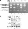
Figure 1.
Figure 1.
(A) RT–PCR analysis of Akt1, Akt2, and Akt3 RNA expression in mouse embryo fibroblasts (MEFs) of different genotypes derived from double-heterozygous matings. Complementary DNA was amplified by PCR with primers specific for mouse Akt1, Akt2, Akt3, and GAPDH (internal control). All PCR products were of the expected size. (Lane 1) Akt1+/+/Akt2+/- MEF. (Lane 2) Akt1-/-/Akt2+/+ MEF. (Lane 3) Akt1-/-/Akt2-/- MEF. (Lane 4) WT MEF. (Lane 5) Akt1+/+/Akt2-/- MEF. (B) Immunoblot showing expression of Akt1 and Akt2 proteins in skeletal muscles of WT and mutant E18.5 embryos, using anti-Akt1 and anti-Akt2. (C) Side views of E18.5 embryos and newborn littermates. WT, wild type; DHet, Akt1+/-/Akt2+/-; DKO, Akt1-/-/Akt2-/-. (D) Newborn body weight (BW) of WT and Akt1/Akt2 mutant mice. The relative average BW (percent of mutant to WT newborns) is shown in parentheses. Results are mean ± S.E.M. *, P < 0.05; ***, P < 0.001.
Figure 1.
Figure 1.
(A) RT–PCR analysis of Akt1, Akt2, and Akt3 RNA expression in mouse embryo fibroblasts (MEFs) of different genotypes derived from double-heterozygous matings. Complementary DNA was amplified by PCR with primers specific for mouse Akt1, Akt2, Akt3, and GAPDH (internal control). All PCR products were of the expected size. (Lane 1) Akt1+/+/Akt2+/- MEF. (Lane 2) Akt1-/-/Akt2+/+ MEF. (Lane 3) Akt1-/-/Akt2-/- MEF. (Lane 4) WT MEF. (Lane 5) Akt1+/+/Akt2-/- MEF. (B) Immunoblot showing expression of Akt1 and Akt2 proteins in skeletal muscles of WT and mutant E18.5 embryos, using anti-Akt1 and anti-Akt2. (C) Side views of E18.5 embryos and newborn littermates. WT, wild type; DHet, Akt1+/-/Akt2+/-; DKO, Akt1-/-/Akt2-/-. (D) Newborn body weight (BW) of WT and Akt1/Akt2 mutant mice. The relative average BW (percent of mutant to WT newborns) is shown in parentheses. Results are mean ± S.E.M. *, P < 0.05; ***, P < 0.001.
Figure 2.
Figure 2.
Skin development is impaired in Akt1/Akt2 DKO mice. (A) Side and top views of newborn WT and Aktl/Akt2 DKO littermates demonstrating the translucent skin of DKO newborns. (B) Histological comparison of the epidermis of newborn littermates. Hematoxylin and eosin (H&E) staining of sections (heart and ling-level cross sections) is shown, C, statum cornetum; G, stratum granulosum; S, stratum spinosum; B, stratum basal; HF, hari follicle. Skin development is impaired in Akt1/Akt2 DKO mice. (C) Analysis of the epidermis from WT and DKO littermates (heart and lung level) with differentiation-specific markets (400× magnification). Paraffin-embedded skin sections were stained with anti-PCNA, K14, K10, or filaggrin antibodies, and detected counterstained with hematoxylin as described in Materials and Methods.
Figure 2.
Figure 2.
Skin development is impaired in Akt1/Akt2 DKO mice. (A) Side and top views of newborn WT and Aktl/Akt2 DKO littermates demonstrating the translucent skin of DKO newborns. (B) Histological comparison of the epidermis of newborn littermates. Hematoxylin and eosin (H&E) staining of sections (heart and ling-level cross sections) is shown, C, statum cornetum; G, stratum granulosum; S, stratum spinosum; B, stratum basal; HF, hari follicle. Skin development is impaired in Akt1/Akt2 DKO mice. (C) Analysis of the epidermis from WT and DKO littermates (heart and lung level) with differentiation-specific markets (400× magnification). Paraffin-embedded skin sections were stained with anti-PCNA, K14, K10, or filaggrin antibodies, and detected counterstained with hematoxylin as described in Materials and Methods.
Figure 3.
Figure 3.
Skeletal muscle atrophy in Akt1/Akt2 DKO mice. (A) Images of standard H&E-stained diaphragms from WT, DHet, and DKO E18.5 littermate embryos (630× magnification). (B) Histograms showing the size and diameter of muscle cells. Digitized images of skeletal muscles were analyzed as described in Materials and Methods. Results are mean ± S.E.M., expressed as the number of pixels in cross-sectional areas of diaphragm and presternum muscle cells or within the width of intercostal muscles. Twenty cells in three random fields were analyzed for each. P < 0.05.
Figure 4.
Figure 4.
mTOR activity in WT and DKO MEFs. (A) Phosphorylation and protein levels were determined by immunoblotting. (Lanes 1,2) Proliferating (P) cells (passage 4) were plated in 10% FCS and analyzed 24 h after plating. (Lanes 3,6) Cells were plated in 10% FCS for 24 h, then deprived of serum and analyzed 24 h later. (Lanes 4,5,7,8) After 24 h of serum deprivation, cells were stimulated with 20% FCS for 30 and 60 min, respectively. Protein extracts from proliferating, serum-deprived, or serum-stimulated cells were subjected to immunoblotting using anti-phospho-Ser 473 of Akt (Akt-p), anti-pan-Akt (Akt-total), anti-phospho-Ser 65 of 4E-BP1 (4E-BP1-p), anti-4E-BP1 (4E-BP1-total), anti-phospho-Thr 389 of S6K1 (S6K1-p), anti-S6K1 (anti-S6K1), or anti-β-actin (loading control). (B, top panel) TSC2 phosphorylation was determined by immunoblotting using anti-phospho-Thr 1452 of TSC2. (Bottom panel) Immunoprecipitation with anti-TSC2 followed by immunoblotting with anti-Akt-p-S/T substrate. ns, nonspecific band. (Lanes 1,4) Cells were plated in 10% FCS for 24 h and then deprived of serum and analyzed 24 h later. (Lanes 2,3,5,6) After 24 h of serum deprivation, cells were stimulated with 20% FCS for 30 and 60 min, prior to analysis.
Figure 5.
Figure 5.
Bone development in WT, DHet, and DKO embryos. (A) Whole skeletons of E14.5 and E18.5 embryos and newborns (NB) were analyzed for ossification using alcian blue and alizarin red. Ossification is visible in the cranium of E14.5 WT and DHet embryos but not in the cranium of E14.5 DKO embryos. Ossification is nearly complete in WT and DHet newborns, whereas large cartilaginous regions are still present in the cranium and skeleton of DKO newborns. (B) Top view of the cranium (top panel) and side view of the hind limb (bottom panel) of WT and DKO newborns showing cartilaginous regions in the interparietal, exoccipital, and surpraoccipital areas (top panel) and in the femur, tibia, and fibula (bottom panel).
Figure 6.
Figure 6.
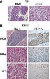
Impaired adipogenesis in Akt1/Akt2 DKO mice. (A) H&E-stained cross sections of the dorsal fat pad at heart level of DHet and DKO newborn littermates (400× magnification). (B) Cross sections of the dorsal fat pad at heart level of E18.5 WT, DHet, and DKO littermates. (Left panels) H&E staining. (Right panels) Staining with anti-PCNA(400× magnification).
Figure 7.
Figure 7.
Akt1/Akt2 DKO MEFs are imparied in adipogenic differentiations and induction of PPARγ. MEFs (passage 4) derived from WT and Akt1/Akt2 DKO embryos were grown to confluence. Two days after confluency (day 0), differentiation was induced with IBMX/DEX/INS. After 8 d of differentiation, cells were observed by light microscopy or stained with Oil-Red-O. (A) Microscopic images of MEFs following induction of adipogenesis. Lipid droplets are clearly visible in WT cells but not in DKO cells. (B) Oil-Red-O staining of differentiating MEFs. (Top panel) Stained plates. (Bottom panel) Microscopic images of stained cells. Akt1/Akt2 DKO MEFs are imparied in adipogenic differentiations and induction of PPARγ. MEFs (passage 4) derived from WT and Akt1/Akt2 DKO embryos were grown to confluence. Two days after confluency (day 0), differentiation was induced with IBMX/DEX/INS. After 8 d of differentiation, cells were observed by light microscopy or stained with Oil-Red-O. (C) Quantification of lipid incorporation by measuring the intensity of Oil-Red-O of WT, Akt1 KO, Akt2 KO, and Akt1/Akt2 DKO MEFs induced to differentiate to adipocytes, presented as average percentage of Oil-Red-O intensity in mutant cells relative to WT cells. The data represent the mean ± S.E.M. from three independent experiments. Significante: p < 0.05, DKO < AKt1 KO < Akt2 KO. There was no significant difference between the Akt2 KO and WT MEFSs. (D) RT-PCR analysis showing PPARγ and C/EBPα mRNA expression in WT and Akt1/Akt2 DKO MEFs after induction of adipocyte differentiation, with GAPDH as an internal control. Days following addition of IBMX/DEX/INS are indicated. (E) RT-PCR analysis showing C/EBPβ, C/EBPδ, and CHOP10 expression following addition of IBMX/DEX/INS. (F) Immunoblots with anti-phospho-Ser 253 of FOXO1 (FOXO1-p) and anti-FOXO1 showing FOXO1 phosphorylation in WT and DKO MEFs following induction of differentiation. (G) Ectopic expression of PPARγ partially restores adipocyte differentiation in DKO MEFs. Cells were infected with either control virus (pBabe) or a PPARγ-expressing retrovirus (pBabe-PPARγ), and were then subjected to differentiation in the absence or presence of 5 μM rosiglitazone (pBabe-PPARγ + rosi), and stained with Oil-Red-O. (H) Schematic illustration, summarizing the molecular mechanism by which Akt may exert its effect on adipocyte differentiation. Akt is required for induction of PPARγ expression during adipocyte differentiation in vitro, and is dispensable for the induction of C/EBPβ and C/EBPδ expression. However, C/EBPβ nd C/EBPδ cannot elicit the induction of PPARγ in the absence of Akt. Akt may exert its effect on PPARγ expression through the forkhead transcription factor FOXO1 or/and through a yet-unknown intermediate factor (see Discussion). Akt may be also required for the induction of C/EBPα expression or, alternatively, PPARγ mediates the effect of Akt on C/EBPα expression
Figure 7.
Figure 7.
Akt1/Akt2 DKO MEFs are imparied in adipogenic differentiations and induction of PPARγ. MEFs (passage 4) derived from WT and Akt1/Akt2 DKO embryos were grown to confluence. Two days after confluency (day 0), differentiation was induced with IBMX/DEX/INS. After 8 d of differentiation, cells were observed by light microscopy or stained with Oil-Red-O. (A) Microscopic images of MEFs following induction of adipogenesis. Lipid droplets are clearly visible in WT cells but not in DKO cells. (B) Oil-Red-O staining of differentiating MEFs. (Top panel) Stained plates. (Bottom panel) Microscopic images of stained cells. Akt1/Akt2 DKO MEFs are imparied in adipogenic differentiations and induction of PPARγ. MEFs (passage 4) derived from WT and Akt1/Akt2 DKO embryos were grown to confluence. Two days after confluency (day 0), differentiation was induced with IBMX/DEX/INS. After 8 d of differentiation, cells were observed by light microscopy or stained with Oil-Red-O. (C) Quantification of lipid incorporation by measuring the intensity of Oil-Red-O of WT, Akt1 KO, Akt2 KO, and Akt1/Akt2 DKO MEFs induced to differentiate to adipocytes, presented as average percentage of Oil-Red-O intensity in mutant cells relative to WT cells. The data represent the mean ± S.E.M. from three independent experiments. Significante: p < 0.05, DKO < AKt1 KO < Akt2 KO. There was no significant difference between the Akt2 KO and WT MEFSs. (D) RT-PCR analysis showing PPARγ and C/EBPα mRNA expression in WT and Akt1/Akt2 DKO MEFs after induction of adipocyte differentiation, with GAPDH as an internal control. Days following addition of IBMX/DEX/INS are indicated. (E) RT-PCR analysis showing C/EBPβ, C/EBPδ, and CHOP10 expression following addition of IBMX/DEX/INS. (F) Immunoblots with anti-phospho-Ser 253 of FOXO1 (FOXO1-p) and anti-FOXO1 showing FOXO1 phosphorylation in WT and DKO MEFs following induction of differentiation. (G) Ectopic expression of PPARγ partially restores adipocyte differentiation in DKO MEFs. Cells were infected with either control virus (pBabe) or a PPARγ-expressing retrovirus (pBabe-PPARγ), and were then subjected to differentiation in the absence or presence of 5 μM rosiglitazone (pBabe-PPARγ + rosi), and stained with Oil-Red-O. (H) Schematic illustration, summarizing the molecular mechanism by which Akt may exert its effect on adipocyte differentiation. Akt is required for induction of PPARγ expression during adipocyte differentiation in vitro, and is dispensable for the induction of C/EBPβ and C/EBPδ expression. However, C/EBPβ nd C/EBPδ cannot elicit the induction of PPARγ in the absence of Akt. Akt may exert its effect on PPARγ expression through the forkhead transcription factor FOXO1 or/and through a yet-unknown intermediate factor (see Discussion). Akt may be also required for the induction of C/EBPα expression or, alternatively, PPARγ mediates the effect of Akt on C/EBPα expression

References

    1. Altomare D.A., Guo, K., Cheng, J.Q., Sonoda, G., Walsh, K., and Testa, J.R. 1995. Cloning, chromosomal localization and expression analysis of the mouse Akt2 oncogene. Oncogene 11: 1055–1060. - PubMed
    1. Altomare D.A., Lyons, G.E., Mitsuuchi, Y., Cheng, J.Q., and Testa, J.R. 1998. Akt2 mRNA is highly expressed in embryonic brown fat and the AKT2 kinase is activated by insulin. Oncogene 116: 2407–2411. - PubMed
    1. Barak Y., Nelson, M.C., Ong, E.S., Jones, Y.Z., Ruiz-Lozano, P., Chien, K.R., Koder, A., and Evans, R.M. 1999. PPAR γ is required for placental, cardiac, and adipose tissue development. Mol. Cell 4: 585–595. - V体育ios版 - PubMed
    1. Bodine S.C., Stitt, T.N., Gonzalez, M., Kline, W.O., Stover, G.L., Bauerlein, R., Zlotchenko, E., Scrimgeour, A., Lawrence, J.C., Glass, D.J., et al. 2001. Akt/mTOR pathway is a crucial regulator of skeletal muscle hypertrophy and can prevent muscle atrophy in vivo. Nat. Cell Biol. 3: 1014–1019. - PubMed
    1. Brazil D.P. and Hemmings, B.A. 2001. Ten years of protein kinase B signalling: Ahard Akt to follow. Trends Biochem. Sci. 26: 657–664. - PubMed

Publication types

MeSH terms

Substances