Skip to main page content (VSports app下载)
U.S. flag

An official website of the United States government

Dot gov

The . gov means it’s official. Federal government websites often end in . gov or . mil. Before sharing sensitive information, make sure you’re on a federal government site. VSports app下载.

Https

The site is secure V体育官网. The https:// ensures that you are connecting to the official website and that any information you provide is encrypted and transmitted securely. .

. 2003 Apr;11(4):1079-92.
doi: 10.1016/s1097-2765(03)00104-7.

A chemical, genetic, and structural analysis of the nuclear bile acid receptor FXR (VSports)

Affiliations

A chemical, genetic, and structural analysis of the nuclear bile acid receptor FXR

V体育ios版 - Michael Downes et al. Mol Cell. 2003 Apr.

"V体育2025版" Abstract

The farnesoid X receptor (FXR) functions as a bile acid (BA) sensor coordinating cholesterol metabolism, lipid homeostasis, and absorption of dietary fats and vitamins. However, BAs are poor reagents for characterizing FXR functions due to multiple receptor independent properties. Accordingly, using combinatorial chemistry we evolved a small molecule agonist termed fexaramine with 100-fold increased affinity relative to natural compounds. Gene-profiling experiments conducted in hepatocytes with FXR-specific fexaramine versus the primary BA chenodeoxycholic acid (CDCA) produced remarkably distinct genomic targets. Highly diffracting cocrystals (1. 78 A) of fexaramine bound to the ligand binding domain of FXR revealed the agonist sequestered in a 726 A(3) hydrophobic cavity and suggest a mechanistic basis for the initial step in the BA signaling pathway. The discovery of fexaramine will allow us to unravel the FXR genetic network from the BA network and selectively manipulate components of the cholesterol pathway that may be useful in treating cholesterol-related human diseases VSports手机版. .


Figures
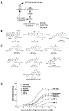
Figure 1.
Figure 1.. Chemical Structures of Identified and Known FXR Ligands and Their In Vitro Binding Affinities
(A) A schematic diagram of the ligand discovery phase and characterization scheme of identified high-affinity agonists. (B) Selected regions of interest for SAR evaluation of prototypical structure lead compounds 1. Region I, right-hand aromatic system; Region II, Acyl group region; Region III, left-hand benzopyran ring system. Compound 2 was produced by systematic optimization of Regions I and II. Fexaramine was selected from a final 94 membered combinatorial library of Region III. (C) Structures of lead compounds (and their EC50 values in a cell-based assay) selected for further biological evaluation as FXR agonists. A, fexaramine (EC50 = 25 nM); B, fexarine (EC50 = 38 nM); C, fexarene (EC50 = 36 nM); D, SRI-1 (EC50 = 377 nM); E, SRI-2 (EC50 = 343 nM). The identified compounds are structurally distinct from known FXR agonists F, CDCA a physiological low-affinity ligand, and the high-affinity ligand G, GW4064 (EC50 = 80 nM). (D) Identified compounds fexaramine, fexarine, fexarene, SRI-1, and SRI-2 are agonists for FXR in vitro. A FRET-based ligand binding assay was carried out in agonist mode using GW4064 as the control ligand. Increasing amounts of the compounds were added as indicated. Binding reactions contained 8 nM europium-labeled GST-FXR-LBD fusion protein and 16 nM allophycocyanin-labeled SRC-1 receptor binding peptide. Results are expressed at 1000*(665 nm/615 nm).
Figure 2.
Figure 2.. Solid Phase Synthesis of a 94 Membered Focused Library of Biaryl and Stilbene Cinnamates
Reagents and conditions: (a) 2.0 equiv of 3, 1.0 equiv of Merrifield Resin (0.91 mmol/g), 2.0 equiv of Cs2CO3, 0.5 equiv of TBAI, DMF, 55°C, 24 hr; (b) 20% TFA in CH2Cl2, 25°C, 1 hr; (c) 10.0 equiv of 4-bromobenzaldehyde, 0.05 equiv of AcOH, THF:MeOH (2:1), 25°C, 1 hr; then, 8.0 equiv of NaCNBH3, THF:MeOH (2:1), 25°C, 2 hr; (d) for R1COCl: 30.0 equiv of R1COCl, 40.0 equiv of Et3N, 1.0 equiv of 4-DMAP, CH2Cl2, 25°C, 12 hr; for R1NCO: 30.0 equiv of R1NCO, 40.0 equiv of Et3N, 1.0 equiv of 4-DMAP, DMF, 65°C, 60 hr; (e) 8.0 equiv of styrene, 10.0 equiv of Et3N, 0.5 equiv of Pd2(dba)3, 1.5 equiv of P(o-tol)3, DMF, 90°C, 48 hr; (f) 5.0 equiv of boronic acid, 3.0 equiv Cs2CO3, 0.5 equiv of Pd(PPh3)4, DMF, 90°C, 24 hr; (g)10.0 equiv of NaOMe, Et2O:MeOH (10:1), 25°C, 20 min. AcOH, acetic acid; 4-DMAP, 4-dimethylaminopyridine; DMF, N,N-dimethylformamide; Et, ethyl; Me, methyl; Pd(PPh3)4, tetrakis(triphenylphosphine)palladium(0); Pd2(dba)3, tris(dibezylideneacetone)dipalladium(0); P(o-tol)3, tri-o-tolylphosphine; TBAI, tetrabutylammonium iodide; TEA, triethylamine; TFA, trifluoroacetic acid; THF, tetrahydrofuran.
Figure 3.
Figure 3.. Characterization of Identified FXR Ligands in Cell-Based Assays
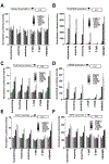
CV-1 cells were cotransfected with pCMX-mFXR and pCMX-hRXR and a reporter gene containing the (A) minimal TK promoter, (B) TK-ECREx6 promoter, (C) TK-ER8×2 promoter, (D) hI-BABP promoter, (E) hPLTP promoter, or (F) rMRP-2 promoter. Increasing amounts (1 nM to 1 μM) of the compounds fexaramine, fexarine, fexarene, SRI-1, SRI-2, and GW4064 were added to the cells 24 hr posttransfection. Activation of the luciferase reporter gene was measured in relative light units with β-galactosidase activity as a control for transfection efficiency and presented as normalized luciferase units. Ligand response data were derived from triplicate points and two independent experiments and presented as the mean ±SE; n = 6.
Figure 4.
Figure 4.. Crossreactivity Studies of the Identified FXR Ligands with Other Nuclear Hormone Receptors
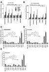
CV-1 cells were cotransfected with a reporter gene containing the (A) MH2004 promoter that contains four GAL4 binding sites with pCMXGAL4-FXR LBD chimeric expression construct or (B) MH2004 promoter with pCMXGAL4-FXR LBD/RXRα constructs and treated with increasing amounts of the compounds fexaramine, fexarine, fexarene, SRI-1, SRI-2, and GW4064. (C, D, and E) CV-1 cells were transiently transfected with the indicated plasmids and treated with either DMSO or 10 μM of the compounds fexaramine, fexarine, and fexarene. Reporter activity was normalized to the internal control, and the data were plotted as fold activation relative to untreated cells. All transfections contained CMX-gal as an internal control.
Figure 5.
Figure 5.. Gene Induction of Identified FXR Targets by Distinct Fexa-Compounds
(A) Total RNA (20 μg) isolated from HT29 stable cells was used for Northern blot analysis. cDNA probes for mFXR and hI-BABP were hybridized to the blot. Blots were normalized to β-actin expression levels. (B and C) HT29 stable cells were then treated overnight with increasing amounts of CDCA (B) and GW4064 (C) as indicated. Total RNA (20 μg) from treated cells was used for Northern blot analysis. cDNA probes for hI-BABP were prepared and hybridized to the blot. Blots were normalized to β -actin. (D) HT29-FXRFL stable cells were cultured until confluence. Cells were then treated overnight with increasing amounts of fexaramine, fexarine, or fexarene as indicated. Total RNA (20 μg) was then isolated using Trizol and used for Northern blot analysis. cDNA probe for human I-BABP was prepared and hybridized to the blot. Blots were normalized to β -actin. (E) HEPG2-FXRFL stable cells were cultured until confluence. Cells were then treated with increasing amounts of fexaramine, fexarine, fexarene, SRI-1, SRI-2, GW4064 (10 nM, 100 nM, 1 μM, 10 μM), and CDCA (1 μM, 10 μM, 100 μM). Twenty micrograms total RNA was then isolated using Trizol and was used for Northern blot analysis. cDNA probes for human PLTP, SHP, MRP-2, and BSEP were prepared and hybridized to the blot. Blots were normalized to 36B4 expression.
Figure 6.
Figure 6.. Gene Array Studies with FXR Ligands Fexaramine, GW4064, and CDCA
(A) Primary mouse hepatocytes were treated for 6 or 12 hr with either vehicle alone or 100 μM CDCA, 10 μM fexaramine, or 10 μM GW4064, as indicated. Total RNA was isolated using Trizol, and 10 μg total RNA was used for Northern blot analysis. The human SHP probe was prepared and hybridized to the blot. To ensure constant loading of total RNA to the blot, GAPDH was also hybridized as a control (data not shown). (B) Clustergram of genes whose expression pattern is altered by FXR agonist treatment. Genes were identified using a paired Student’s t test and DMSO treatment as the control group. Transcripts (222) were identified meeting a criteria of a change of at least 0.005 and a ≥2-fold change with respect to DMSO. Data was imported into the cluster, and the genes were subjected to hierarchal clustering. The output was visualized using Treeview. Red coding indicates induction relative to other conditions, green indicates repression, and black indicates no change. (C) Table of genes that were commonly repressed or activated in primary human hepatocytes by CDCA, fexaramine, or GW4064 compared with DMSO treatment. Fold changes indicated represent the average change over the three treatments. – indicates repressed gene message while + indicates an increase in gene expression.
Figure 7.
Figure 7.. Crystal Structure of FXR Bound to Fexaramine
(A) Structure of hFXR-LBD. Residues 248 to 270 and 286 to 476 of hFXR-LBD in complex with the high-affinity agonist fexaramine. The α1 helices are shown in blue, and the ligand is shown in gold embedded in a transparent van der Waals surface. The structural elements are numbered according to the canonical structure for the LBD of nuclear receptors (NSB ref for canonical label). (B) Sequence alignment of FXR, VDR, SXR, and RXRα1 LBDs. The secondary structural elements of the hFXR-LBD are shown above the FXR sequence in blue and are labeled accordingly (see Figure 1A). Hydrophobic residues involved in binding fexaramine are highlighted in violet. Polar interactions are shown in blue and red. (C) Close-up of the first set of interactions with fexaramine. The hexyl group protrudes out into solution while making weak van der Waals contact with I339 and L344. The fexaramine carbonyl oxygen participates in two hydrogen bonding interactions (H298 and S336). The methyl ester aliphatic chain makes van der Waals contacts with Met294, Leu352, and I356. No charged interactions are seen in contact with the methyl ester moiety itself. (D) Close-up of the second set of interactions with fexaramine. The double benzyl rings make van der Waals contact with 15 residues. The majority of the ligand binding pocket is hydrophobic and partially aromatic in nature. (E) Close-up of a proposed model for complexation of CDCA by FXR-LBD. CDCA was modeled on the experimentally derived orientation of fexaramine. CDCA’s two hydroxyl groups are pointed toward the side chains of Y365 and H451 to putatively participate in favorable hydrogen bonding. This positions the CDCA carboxyl group in the same orientation as the fexaramine hexyl group, suggesting that it protrudes from the protein or makes contacts with the insertion domain region. Notably, glycine and taurine bile acid conjugates could be accommodated in this orientation, which affords steric accommodation of the cognate tails.

"V体育ios版" Comment in

References (V体育平台登录)

    1. Blumberg B, and Evans RM (1998). Orphan nuclear receptors—new ligands and new possibilities. Genes Dev. 12, 3149–3155. - PubMed
    1. Blumberg B, Sabbagh W Jr., Juguilon H, Bolado J Jr., van Meter CM, Ong ES, and Evans RM (1998). SXR, a novel steroid and xenobiotic-sensing nuclear receptor. Genes Dev. 12, 3195–3205. - PMC - PubMed
    1. Brünger AT, Adams PD, Clore GM, DeLano WL, Gros P, Grosse-Kunstleve RW, Jiang JS, Kuszewski J, Nilges M, Pannu NS, et al. (1998). Crystallography & NMR system: A new software suite for macromolecular structure determination. Acta Crystallogr. D Biol. Crystallogr. 54, 905–921. - "V体育ios版" PubMed
    1. Chawla A, Saez E, and Evans RM. (2000). “Don’t know much bile- ology”. Cell. 103, 1–4. - VSports app下载 - PubMed
    1. Chiang JY (2002). Bile acid regulation of gene expression: roles of nuclear hormone receptors. Endocr. Rev. 23, 443–463. - PubMed

Publication types

MeSH terms

V体育官网 - Associated data