Skip to main page content (VSports注册入口)
U.S. flag

An official website of the United States government

Dot gov

The VSports app下载. gov means it’s official. Federal government websites often end in . gov or . mil. Before sharing sensitive information, make sure you’re on a federal government site. .

Https

The site is secure. The https:// ensures that you are connecting to the official website and that any information you provide is encrypted and transmitted securely V体育官网. .

. 2002 Jun 1;30(11):2349-57.
doi: 10.1093/nar/30.11.2349.

Human OGG1 undergoes serine phosphorylation and associates with the nuclear matrix and mitotic chromatin in vivo

Affiliations

Human OGG1 undergoes serine phosphorylation and associates with the nuclear matrix and mitotic chromatin in vivo (VSports在线直播)

Françoise Dantzer et al. Nucleic Acids Res. .

Abstract

OGG1 is the major DNA glycosylase in human cells for removal of 7,8 dihydro-8-oxoguanine (8-oxoG), one of the most frequent endogenous base lesions formed in the DNA of aerobic organisms. During replication, 8-oxoG will frequently mispair with adenine, thus forming G:C --> T:A transversions, a common somatic mutation associated with human cancers. In the present study, we have constructed a stable transfectant cell line expressing hOGG1 fused at the C-terminal end to green fluorescent protein (GFP) and investigated the cellular distribution of the fusion protein by fluorescence analysis. It is shown that hOGG1 is preferentially associated with chromatin and the nuclear matrix during interphase and becomes associated with the condensed chromatin during mitosis. Chromatin-bound hOGG1 was found to be phosphorylated on a serine residue in vivo as revealed by staining with an anti-phosphoserine-specific antibody. Chromatin-associated hOGG1 was co-precipitated with an antibody against protein kinase C (PKC), suggesting that PKC is responsible for the phosphorylation event. Both purified and nuclear matrix-associated hOGG1 were shown to be substrates for PKC-mediated phosphorylation in vitro. This appears to be the first demonstration of a post-translational modification of hOGG1 in vivo VSports手机版. .


Figures
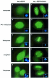
Figure 1
Figure 1
Subcellular distribution of hOGG1. HeLa cells expressing EGFP (AE) or the hOGG1–EGFP fusion protein (FJ) were grown on coverslips, fixed and analysed using an Axioplan 2 fluorescence microscope (Carl Zeiss). DNA (inserts) was labelled with DAPI staining.
Figure 2
Figure 2
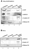
hOGG1 is associated with the chromatin and the nuclear matrix. HeLa cells expressing the hOGG1–EGFP fusion protein (A) or non-transfected HeLa cells (B) were extracted to obtain the cytoplasmic fraction (lane 1), the whole chromatin fraction (lane 2), a high salt wash (lane 3) and the core nuclear matrix (lane 4). Proteins from equal cell equivalents from each fraction were analysed by western blotting with the indicated antibodies.
Figure 3
Figure 3
In situ extraction of the core nuclear matrix. HeLa cells expressing hOGG1–EGFP (ac) or EGFP (df) were grown on coverslips, extracted sequentially as described in Figure 2, fixed and processed for immunofluorescence imaging. (a and d) The validity of the procedure is shown by the absence of DAPI-stained nuclear DNA. (b and e) GFP (green) and (c and f) Alexa 594-labelled lamin A/C.
Figure 4
Figure 4
Comparative 8-oxoG cleavage activity in chromatin and nuclear matrix fractions. (A) Schematic representation of hOGG1 enzymatic activity. A 24 bp double-stranded oligonucleotide containing an 8-oxoG:C base pair at position 14 (in bold) was prepared as described (13) and 32P-labelled at the 5′ end of the 8-oxoG-containing strand. hOGG1 activity excises the 8-oxoG and cleaves the 32P-labelled 8-oxoG-containing strand 3′ of the lesion (β-elimination). The addition of HAP1 endonuclease induces subsequent cleavage of the terminal sugar phosphate. Both lyase and endonuclease activities produce a 13 nt 32P-end-labelled product when the reaction is run on denaturating polyacrylamide gels. (B) HeLa cells expressing hOGG1–EGFP or (C) non-transfected HeLa cells were fractionated as described in the Material and Methods and shown in Figure 2. Equivalent amounts of each fraction were incubated with the 8-oxoG:C-containing double-stranded oligonucleotide. Arrows indicate the positions of the 5′ radiolabelled substrate (24 bp) and reaction products as follows: 13 nt, 13mer product produced by hOGG1 strand nicking activity (lanes 1–4); 13* nt, 13 mer with a 3′-OH produced by the addition of HAP1 endonuclease where indicated (lanes 5–8). Ten nanograms of purified hOGG1 was added in control reaction mixtures with (lane 9) or without (lane 10) nuclear matrix extract. Cleavage products were analysed by PhosphorImaging after 20% denaturating PAGE.
Figure 5
Figure 5
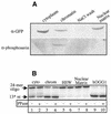
The chromatin-associated hOGG1–EGFP is phosphorylated on a serine residue in vivo. (A) HeLa cells expressing the hOGG1–EGFP fusion protein were extracted to obtain the cytoplasmic fraction, the whole chromatin fraction, a high salt wash and the core nuclear matrix. Proteins from equal cell equivalents of each fraction were analysed by western blotting with the indicated antibodies. (B) Equal cell equivalents from each fraction (lanes 1–8) or 10 ng of hOGG1 (lanes 9 and 10) were mock treated (lanes 1, 3, 5, 7 and 9) or treated with λ-PPase (lanes 2, 4, 6, 8 and 10) before measuring hOGG1 activity in the presence of HAP1 endonuclease as described in Figure 4. 13* nt, 13 mer with a 3′-OH produced by the addition of HAP1 after hOGG1-strand nicking activity.
Figure 6
Figure 6
The chromatin-associated hOGG1–EGFP is co-immunoprecipitated with PKC in vivo. Equal cell equivalents from HeLa hOGG1–EGFP (A) or HeLa EGFP (B) biochemical fractions were subjected to immunoprecipitation with either anti-GFP or anti-PKC antibody as indicated. Immunoprecipitates were then analysed by immunoblotting with the appropriate antibodies.
Figure 7
Figure 7
PKC phosphorylates purified hOGG1 and the nuclear matrix-associated hOGG1–EGFP in vitro. (A) Purified recombinant hOGG1 protein was incubated in the presence (lanes 1 and 2) or in the absence (lane 3) of PKC together with [γ-32P]ATP (added in lanes 1 and 3) (left panel). The position of the phosphorylated protein was identified by comparing with purified hOGG1 run on a Coomassie-stained gel (lane 4, right panel). An equivalent of 10 ng of protein was assayed for hOGG1 enzymatic activity as described in Figure 4 (lower panel). (B) Cell extract equivalents from chromatin (lanes 1 and 3) and nuclear matrix fractions (lanes 2 and 4) from HeLa hOGG1–EGFP cells were incubated in the presence (lanes 3 and 4) or in the absence (lanes 1 and 2) of PKC together with [γ-32P]ATP (left panel). The position of phosphorylated hOGG1–EGFP was identified by Coomassie staining of the treated fraction (right panel). Following the phosphorylation reaction, samples were assayed for hOGG1 enzymatic activity as described in Figure 4 (lower panel).

References

    1. Marnett L.J. (2000) Oxyradicals and DNA damage. Carcinogenesis, 21, 361–370. - PubMed
    1. Raha S. and Robinson,B.H. (2000) Mitochondria, oxygen free radicals diseases and ageing. Trends Biochem. Sci., 25, 502–508. - PubMed (V体育ios版)
    1. Seeberg E., Eide,L. and Bjørås,M. (1995) The base excision repair pathway. Trends Biochem. Sci., 20, 391–397. - PubMed
    1. Cheng K.C., Cahill,D.S., Kasai,H., Nishimura,S. and Loeb,L.A. (1992) 8-Hydroxyguanine, an abundant form of oxidative DNA damage, causes G-T and A-C substitutions. J. Biol. Chem. 267, 166–172. - PubMed
    1. Moriya M. (1993) Single-stranded shuttle phagemid for mutagenesis studies in mammalian cells: 8-oxoguanine in DNA induces targeted G.C → T.A transversions in simian kidney cells. Proc. Natl Acad. Sci. USA, 90, 1122–1126. - "V体育平台登录" PMC - PubMed

Publication types

MeSH terms (VSports最新版本)