"VSports手机版" Skip to main page content
U.S. flag

An official website of the United States government

Dot gov

The . gov means it’s official VSports app下载. Federal government websites often end in . gov or . mil. Before sharing sensitive information, make sure you’re on a federal government site. .

Https

The site is secure V体育官网. The https:// ensures that you are connecting to the official website and that any information you provide is encrypted and transmitted securely. .

. 2001 Mar 30;276(13):10374-86.
doi: 10.1074/jbc.M010271200. Epub 2000 Dec 28.

A conserved docking site in MEKs mediates high-affinity binding to MAP kinases and cooperates with a scaffold protein to enhance signal transmission

Affiliations

"VSports手机版" A conserved docking site in MEKs mediates high-affinity binding to MAP kinases and cooperates with a scaffold protein to enhance signal transmission

A J Bardwell et al. J Biol Chem. .

Abstract

The recognition of mitogen-activated protein kinases (MAPKs) by their upstream activators, MAPK/ERK kinases (MEKs), is crucial for the effective and accurate transmission of many signals. We demonstrated previously that the yeast MAPKs Kss1 and Fus3 bind with high affinity to the N terminus of the MEK Ste7, and proposed that a conserved motif in Ste7, the MAPK-docking site, mediates this interaction. Here we show that the corresponding sequences in human MEK1 and MEK2 are necessary and sufficient for the direct binding of the MAPKs ERK1 and ERK2. Mutations in MEK1, MEK2, or Ste7 that altered conserved residues in the docking site diminished binding of the cognate MAPKs. Furthermore, short peptides corresponding to the docking sites in these MEKs inhibited MEK1-mediated phosphorylation of ERK2 in vitro. In yeast cells, docking-defective alleles of Ste7 were modestly compromised in their ability to transmit the mating pheromone signal. This deficiency was dramatically enhanced when the ability of the Ste5 scaffold protein to associate with components of the MAPK cascade was also compromised. Thus, both the MEK-MAPK docking interaction and binding to the Ste5 scaffold make mutually reinforcing contributions to the efficiency of signaling by this MAPK cascade in vivo VSports手机版. .


Figures
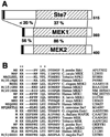
FIG. 1
FIG. 1. A MAPK-docking site near the N terminus of MEKs
A, schematic representation of S. cerevisiae Ste7, and human MEK1 and MEK2, showing the positions of the MAPK-docking site (solid bar) and the protein kinase catalytic core (hatched region). The percent amino acid identity between Ste7 and MEK1 and between MEK1 and MEK2 is indicated for the N-terminal and catalytic domains. Relatedness between Ste7 and MEK2 (<20 and 36% identity, respectively, in the N-terminal and catalytic domains) is similar to that between Ste7 and MEK1. Length of each protein is indicated on the right. B, alignment of the N termini of representatives of the MEK/MKK/MAPKK family of dual-specificity protein kinases. All sequences start from the initiator methionine. Positions of the doublet of basic residues and the nonpolar residues of the hydrophobic-X-hydrophobic element of the core MAPK-docking site motif are indicated by asterisks. One-residue gaps are indicated by hyphens (−); spaces are for visual clarity. Source of the sequence shown is given on the right. Accession numbers for the NCBI Nucleotide or Protein data bases are given. In most cases, rodent and other mammalian MEKs are identical to their human orthologs in the docking-site region; hence they are not shown to reduce redundancy. MEKs containing an N-terminal sequence that nearly fits the above consensus (e.g. Drosophila Dsor1 (Q24324) and p38 MAPKK (CAB45101), and Leishmania MAPKK (CAB45417)) are also not shown.
FIG. 2
FIG. 2. The MAPK-docking site is sufficient for MAPK binding
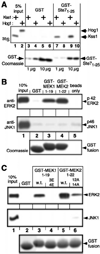
A, Kss1, but not Hog1, binds to the MAPK-docking site of Ste7. Radiolabeled (35S) Kss1 and Hog1 proteins were prepared by in vitro translation, partially purified by ammonium sulfate precipitation, and portions (5% of the amount added in the binding reactions; input) were resolved on a 10% SDS-polyacrylamide gel (lanes 1 and 2). Samples (1 pmol) of the same proteins were incubated with 1 or 10 µg of GST (lanes 3–6) or GST-Ste71–25 (lanes 7–10), bound to glutathione-Sepharose (GSH) beads, and the resulting bead-bound protein complexes were isolated by sedimentation and resolved by 10% SDS-PAGE on the same gel. The gel was analyzed by staining with Coomassie Blue for visualization of the bound GST fusion protein (lower panel), and by autoradiography for visualization of the bound radiolabeled protein (upper panel). B, ERK2, but not JNK1, binds to the MAPK-docking sites of MEK1 and MEK2. Purified ERK2 or JNK1α1 proteins (1 pmol) were incubated with ~100 µg of GST, GST-MEK11–19, or GST-MEK21–22 that were pre-bound to GSH beads (lanes 2–4), or with empty GSH beads only (lane 5). Bead-bound complexes were isolated, resolved by 10% SDS-PAGE, and analyzed by staining with Coomassie Blue for visualization of the bound GST fusion protein (lower panel), and by immunostaining with anti-ERK2 (upper panel) or anti-JNK1 (middle panel) antisera. Input (10% of the amount added in the binding reactions) is shown in lane 1. The middle panel is overexposed to highlight that there was no binding of JNK1 above background. C, alteration of conserved residues in the MAPK-docking sites of MEK1 and MEK2 result in diminished MAPK binding. Experimental design as in B, except that the GST fusion proteins were GST, GST-MEK11–19, GST-MEK11–19EE, GST-MEK21–22, and GST-MEK21–22ATA (lanes 2–6, respectively).
FIG. 3
FIG. 3. High-affinity interaction between full-length MEK and ERK requires the MAPK-docking site
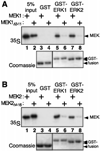
A, MEK1, but not MEK1Δ3–11, binds to ERK1 and ERK2. Radiolabeled (35S) MEK1 and MEK1Δ3–11 proteins (~1 pmol) were incubated with 10 µg of purified GST, GST-ERK1, or GST-ERK2 that were pre-bound to GSH beads, and bead-bound protein complexes were isolated and analyzed as in Fig. 2A. Inputs (5% of the amount added in the binding reactions) are shown in lanes 1 and 2. B, MEK2, but not MEK2Δ4–16, binds to ERK1 and ERK2. Experimental design as in A, except that the radiolabeled proteins were MEK2 and MEK2Δ4–16, as indicated.
FIG. 4
FIG. 4. Conserved residues in the MAPK-docking site of Ste7 are required for high-affinity MAPK binding
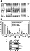
A, Ste7 MAPK-docking site mutants analyzed. The first 27 residues of Ste7 are shown. The positions of substitution mutations are shown in bold and underlined. Residues removed in deletion mutations are indicated by hyphens (−); spaces are for visual clarity. The name of the mutant allele is on the left; a summary of the binding data, shown in B is on the right. B, relative binding of radiolabeled Ste7N (either Ste71–172 or Ste71–98) and mutant derivatives thereof to GST-Fus3 (solid bars) or to GST-ERK2 (open bars). 35S-Ste7N and mutants thereof (2 pmol) were incubated with 20 pmol of GST, 2 pmol of GST-Fus3, or 2 pmol of GST-ERK2 bound to GSH beads, and the resulting bead-bound protein complexes were quantified in a scintillation counter. The nonspecific background adsorption of each radiolabel protein to GST alone was subtracted. Under these conditions, 8.1% of the input wild-type Ste7N bound to GST-Fus3, and 4.7% bound to GST-ERK2. Results were normalized by setting these values as 100%. Data shown are the average of at least three experiments; error bars indicate standard deviations. The relative affinity of the weak-binding derivatives was confirmed in experiments using 20 pmol of GST-Fus3 (not shown). C, representative data for experiments shown above. Radiolabeled wild-type Ste7N, or the AAAA or Δ2–7 derivatives, as indicated, was incubated with 20 pmol of GST (G; lane 2), or with 2 or 20 pmol of GST-Fus3 (lanes 3 and 4, respectively) that had been pre-bound to GSH beads. Bead-bound complexes were isolated, resolved by 12% SDS-PAGE, and visualized by autoradiography. Input (10% of the amount used in the binding reactions) is shown in lane 1.
FIG. 5
FIG. 5. Inhibition of MEK1-dependent phosphorylation of ERK2 by MAPK-docking site peptides
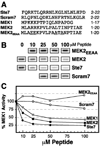
A, peptides used. Sequences of the synthetic peptides prepared for this study are shown; substitution mutations are indicated in bold and underlined. The corresponding residues of the full-length proteins are given on the right. B and C, effect of peptides on phosphorylation of ERK2 by MEK1. Purified, full-length, catalytically inactive ERK2 (1.25 µm) was incubated with 1 unit of purified active MEK1 and [γ-32P]ATP for 20 min, in the absence or presence of the specified concentrations of the indicated peptides. ERK2 phosphorylation was quantified by SDS-PAGE followed by analysis of relative incorporation using a PhosphorImager™ (Molecular Dynamics, Inc.). B, autoradiogram of a representative experiment. C, results plotted as percent phosphorylation relative to that observed in the absence of any added peptide. Data are the average of at least three experiments.
FIG. 6
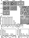
FIG. 6. Overlap between the MAPK docking function of Ste7 and the scaffold function of Ste5
A, synergistic effect of scaffolding-deficient Ste5 and docking-defective Ste7 on pheromone-imposed cell-cycle arrest. Yeast strain YLB105 (MATa ste5Δ ste7Δ) was transformed with a plasmid encoding wild-type Ste5, or Ste5D746G, or an “empty” vector, in combination with a plasmid encoding wild-type Ste7, Ste7Δ2–19, or an empty vector. The resulting transformants were plated as a lawn, onto which were placed discs containing 12 µg (left) or 3 µg (right) of α-factor mating pheromone. The plates were photographed 48 h later. Pheromone-imposed cell-cycle arrest is indicated by the zone of growth inhibition (“halo”) surrounding a disc. B and C, scaffolding-defective Ste5 unmasks a role for the Ste7-MAPK docking interaction in mating proficiency. Strain YLB105 was transformed with a plasmid encoding wild-type Ste5 or Ste5V763A/S861P (denoted VASP in B), in combination with a plasmid encoding wild-type Ste7, or mutant derivatives thereof. B, the resulting transformants were streaked onto a plate selective for plasmid maintenance (top), then scored for mating to an appropriate tester strain using a qualitative assay (bottom). C, the resulting transformants were scored for mating to an appropriate tester strain using a quantitative assay. Mating efficiencies (diploids formed per input MATa haploid) are normalized to the value (0.51) obtained for cells expressing wild-type Ste5 and wild-type Ste7. Error bars indicate standard deviations of duplicate experiments. D and E, partially redundant roles for scaffolding and docking in transmitting a signal from constitutively active MEKK (Ste11). Strain YLB105 was transformed with a plasmid encoding wild-type Ste5 or an empty vector (D and E), or Ste5V763A/S861P (E), in combination with a plasmid encoding wild-type Ste7, or mutant derivatives thereof. The resulting transformants were then further transformed with a plasmid containing a reporter gene regulated by the mating MAPK cascade (FUS1-lacZ), and a plasmid carrying dominant, constitutively active STE11-4. After 4 days, the resulting colonies were pooled, and expression of the reporter gene was assessed by determination of β-galactosidase specific activity. Values are normalized to that obtained for cells expressing wild-type Ste5 and wild-type Ste7, and represent the average of at least three experiments. Error bars indicate standard deviations.
FIG. 7
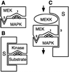
FIG. 7. Diagrammatic representation of interactions between MEKs, MAPKs and scaffold proteins
A, the ability of a MEK to interact with its cognate MAPK both by an active site-target residue interaction (1) and by a docking interaction (2) comprises, in principle, a “double selection” that has the potential to promote the efficiency and fidelity of MAPK activation. To aid conceptualization, these two interactions are shown as occurring simultaneously. Alternatively, they may occur in kinetically distinct steps (45, 85). The region of the MAPK that is phosphorylated by the MEK is represented by a chain of five circles. B, view of scaffolding as special case of docking (or visa versa). Compare with A, the scaffold protein is indicated by an S. C, overlapping roles of docking and scaffolding in promoting MAPK cascade signaling. MAPK activation is promoted by direct MEK-MAPK binding via the active site (1) and the docking site (2), and by binding of both MEK and MAPK to a scaffold (S) protein (3). In support of the scheme shown, it is known that the catalytic domain of Ste7 binds to Ste5 (27, 29), whereas the N terminus of Ste7 binds to Fus3 (this study and Refs. and 45). It is likely, therefore, that Ste7 can bind to both Ste5 and Fus3 simultaneously. We have shown here that mutations in the MAPK-docking site of MEK (2), combined with mutations in the MEK-binding site (3) of the scaffold Ste5, or with deletion of the scaffold entirely, leads to a synergistic reduction in MAPK activation in the yeast mating pheromone response pathway.

References

    1. Graves JD, Krebs EG. Pharmacol. Ther. 1999;82:111–121. - PubMed
    1. Hunter T. Cell. 2000;100:113–127. - PubMed
    1. Schaeffer HJ, Weber MJ. Mol. Cell. Biol. 1999;19:2435–2444. - PMC - PubMed
    1. Lewis TS, Shapiro PS, Ahn NG. Adv. Cancer Res. 1998;74:49–139. - VSports手机版 - PubMed
    1. Garrington T, Johnson G. Curr. Opin. Cell Biol. 1999;11:211–218. - "VSports注册入口" PubMed

MeSH terms

LinkOut - more resources