"VSports" Physiological expression of the gene for PrP-like protein, PrPLP/Dpl, by brain endothelial cells and its ectopic expression in neurons of PrP-deficient mice ataxic due to Purkinje cell degeneration
- PMID: 11073804
- PMCID: "V体育2025版" PMC1885740
- DOI: 10.1016/S0002-9440(10)64782-7
Physiological expression of the gene for PrP-like protein, PrPLP/Dpl, by brain endothelial cells and its ectopic expression in neurons of PrP-deficient mice ataxic due to Purkinje cell degeneration
Abstract
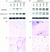
Recently, a novel gene encoding a prion protein (PrP)-like glycoprotein, PrPLP/Dpl, was identified as being expressed ectopically by neurons of the ataxic PrP-deficient (PRNP(-/-)) mouse lines exhibiting Purkinje cell degeneration. In adult wild-type mice, PrPLP/Dpl mRNA was physiologically expressed at a high level by testis and heart, but was barely detectable in brain. However, transient expression of PrPLP/Dpl mRNA was detectable by Northern blotting in the brain of neonatal wild-type mice, showing maximal expression around 1 week after birth. In situ hybridization paired with immunohistochemistry using anti-factor VIII serum identified brain endothelial cells as expressing the transcripts. Moreover, in the neonatal wild-type mice, the PrPLP/Dpl mRNA colocalized with factor VIII immunoreactivities in spleen and was detectable on capillaries in lamina propria mucosa of gut. These findings suggested a role of PrPLP/Dpl in angiogenesis, in particular blood-brain barrier maturation in the central nervous system. Even in the ataxic Ngsk PRNP(-/-) mice, the physiological regulation of PrPLP/Dpl mRNA expression in brain endothelial cells was still preserved. This strongly supports the argument that the ectopic expression of PrPLP/Dpl in neurons, but not deregulation of its physiological expression in endothelial cells, is involved in the neuronal degeneration in ataxic PRNP(-/-) mice VSports手机版. .
Figures


References (VSports app下载)
-
- Prusiner SB: Molecular biology of prion diseases. Science 1991, 252:1515-1522 - PubMed
-
- Büeler H, Fischer M, Lang Y, Bluethmann H, Lipp HP, DeArmond SJ, Prusiner SB, Aguet M, Weissmann C: Normal development and behaviour of mice lacking the neuronal cell-surface PrP protein. Nature 1992, 356:577-582 - PubMed
-
- Manson JC, Clarke AR, Hooper ML, Aitchison L, McConnell I, Hope J: 129/Ola mice carrying a null mutation in PrP that abolishes mRNA production are developmentally normal. Mol Neurobiol 1994, 8:121-127 - VSports app下载 - PubMed
-
- Sakaguchi S, Katamine S, Nishida N, Moriuchi R, Shigematsu K, Sugimoto T, Nakatani A, Kataoka Y, Houtani T, Shirabe S, Okada H, Hasegawa S, Miyamoto T, Noda T: Loss of cerebellar Purkinje cells in aged mice homozygous for a disrupted PrP gene. Nature 1996, 380:528-531 - PubMed
-
- Moore RC, Lee IY, Silverman GL, Harrison PM, Strome R, Heinrich C, Karunaratne A, Pasternak SH, Chishti MA, Liang Y, Mastrangelo P, Wang K, Smit AFA, Katamine S, Carlson GA, Cohen FE, Prusiner SB, Melton DW, Tremblay P, Hood LE, Westaway D: Ataxia in prion protein (PrP) deficient mice associated with upregulation of the novel PrP-like protein doppel. J Mol Biol 1999, 292:797-817 - PubMed
V体育官网 - Publication types
- V体育官网入口 - Actions
MeSH terms
- Actions (V体育平台登录)
- "VSports app下载" Actions
- Actions (V体育官网)
- VSports - Actions
- "VSports在线直播" Actions
- Actions (V体育平台登录)
- V体育安卓版 - Actions
- "V体育2025版" Actions
- "VSports app下载" Actions
Substances
- V体育2025版 - Actions
- "V体育2025版" Actions
- "VSports app下载" Actions
LinkOut - more resources
V体育2025版 - Full Text Sources
VSports app下载 - Other Literature Sources
Molecular Biology Databases
Research Materials

