VSports - Skip to main page content
U.S. flag

An official website of the United States government

Dot gov

The VSports app下载. gov means it’s official. Federal government websites often end in . gov or . mil. Before sharing sensitive information, make sure you’re on a federal government site. .

Https

The site is secure. The https:// ensures that you are connecting to the official website and that any information you provide is encrypted and transmitted securely. V体育官网.

. 2000 Aug 15;97(17):9624-9.
doi: 10.1073/pnas.97.17.9624.

Smad4/DPC4-mediated tumor suppression through suppression of angiogenesis

Affiliations

"VSports" Smad4/DPC4-mediated tumor suppression through suppression of angiogenesis

I Schwarte-Waldhoff et al. Proc Natl Acad Sci U S A. .

Abstract

Smad4/DPC4 (deleted in pancreatic carcinoma, locus 4) is a tumor suppressor gene lost at high frequency in cancers of the pancreas and other gastrointestinal organs. Smad4 encodes a key intracellular messenger in the transforming growth factor beta (TGF-beta) signaling cascade. TGF-beta is a potent inhibitor of the growth of epithelial cells; thus, it has been assumed that loss of Smad4 during tumor progression relieves this inhibition. Herein, we show that restoration of Smad4 to human pancreatic carcinoma cells suppressed tumor formation in vivo, yet it did not restore sensitivity to TGF-beta. Rather, Smad4 restoration influenced angiogenesis, decreasing expression of vascular endothelial growth factor and increasing expression of thrombospondin-1. In contrast to the parental cell line and to control transfectants that produced rapidly growing tumors in vivo, Smad4 revertants induced small nonprogressive tumors with reduced vascular density. These data define the control of an angiogenic switch as an alternative, previously unknown mechanism of tumor suppression for Smad4 and identify the angiogenic mediators vascular endothelial growth factor and thrombospondin-1 as key target genes VSports手机版. .


Figures

Figure 1
Figure 1
Reexpression of the human Smad4/DPC4 in the human pancreatic adenocarcinoma cell line Hs766T. (A) Northern blot analysis of total RNA from the parental cell line and stable transfectants hybridized with a human Smad4/DPC4 cDNA probe. Lane 2, Hs766T parental cell line; lanes 3–7, Smad4/DPC4 reconstituted clones D2, D4, D5, D8, and D9; lanes 8 and 9, negative control clones K3 and K6. (B) Western blot analysis for the human Smad4/DPC4 protein on total protein extracts. Lane 1, human pancreatic adenocarcinoma cell line Paca44 as control for endogenous Smad4/DPC4; lane 2, Hs766T parental cell line; lanes 3–7, Smad4/DPC4 reconstituted clones; lanes 8 and 9, negative control clones.
Figure 2
Figure 2
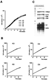
Analysis of growth and TGF-β response in vitro and expression analysis of TGF-β receptors. (A) Growth of Smad4/DPC4-reexpressing cell clones and negative controls cultured in serum-supplemented growth medium. (B) Growth of Smad4/DPC4-reexpressing cells and negative control cells incubated in the absence (continuous bar) or presence (hatched bar) of TGF-β1 (5 ng/ml) under reduced serum concentrations. (A and B) Cells were plated in duplicate (A) or triplicate (B) for each time point, and results were confirmed in at least two independent experiments. Note that error bars for standard deviations are shown in B but are too narrow to be resolved. (C) Expression of TGF-βRI and TGF-βRII as analyzed by RNase protection assay. Lane 1, probe; lane 2, tRNA; lane 3, Hs766T parental cell line; lanes 4 and 5, Smad4/DPC4 negative control clones K3 and K6; lanes 6 and 7, Smad4/DPC4-reconstituted clones D5 and D8.
Figure 3
Figure 3
Suppression of tumor growth in vivo. (A) Mean mass of tumors derived from control clones K3 and K6 and Smad4/DPC4-reexpressing clones D4, D5, and D8 (mean of eight tumors each). All mice were killed, and tumors were prepared and weighed by 17 days after s.c. injection, when tumors from the control clones reached 10 mm in diameter. (B) Time course of tumor growth derived from control clones and Smad4/DPC4-reexpressing clones in an independent experiment (tumor volume = length × width2/2, mean of six tumors from each clone).
Figure 4
Figure 4
Smad4/DPC4-mediated shifts in VEGF and TSP-1 expression levels. (A) Northern blot with total RNA was hybridized with a human VEGF cDNA probe, stripped, and rehybridized with a TSP-1-specific cDNA probe. Lane 1, Hs766T parental cell line; lanes 2 and 3, Smad4/DPC4-negative clones K3 and K6; lanes 4–6, Smad4/DPC4-reconstituted clones D4, D5, and D8. The difference in expression levels was confirmed with at least three independent RNA preparations each. Quantification of RNA levels repeatedly revealed a reduction of the VEGF steady-state mRNA level by a factor of two to three and induction of TSP-1 expression by a factor of approximately three in Smad4/DPC4-reexpressing clones grown in full medium or incubated in serum-free medium. (B) VEGF and TSP-1 Western blots with protein from conditioned media. Lanes 2 and 3, Smad4/DPC4-negative clones K3 and K6; lanes 5 and 6, Smad4/DPC4-reconstituted clones D5 and D8. The signals correspond to dimeric VEGF165 and trimeric TSP-1.
Figure 5
Figure 5
Activation of endothelial cell migration by Smad4/DPC4-negative control cells is due to VEGF, and inhibitory activity of Smad4/DPC4-reconstituted cells is due to TSP-1. (A) Conditioned media (20 μg/ml) were tested to evaluate induction of migration in Smad4/DPC4-negative and Smad4/DPC4-positive cells. (B) Conditioned media from Smad4/DPC4-negative cells were tested in the absence or presence of neutralizing anti-VEGF antibody (at 5 μg/ml). (C) Conditioned media from Smad4/DPC4-positive cells were tested in the absence or presence of neutralizing anti-TSP-1 antibody (20 μg/ml) with or without addition of recombinant VEGF (0.1 ng/ml). (D) Controls demonstrating the effect of neutralizing antibodies and recombinant proteins. All samples were tested in quadruplicate. Bars, standard errors converted to percentages. All experiments were repeated and gave virtually identical results.
Figure 6
Figure 6
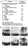
In vivo angiogenic activity of Smad4/DPC4-reconstituted clones and negative control. (A) Hydron pellets containing conditioned medium (200 μg/ml) alone or with inducer (1 ng/ml VEGF or 100 ng/ml basic fibroblast growth factor) or neutralizing antibody to VEGF (5 μg/ml) or TSP-1 (antibody A4.1 at 20 μg/ml) were implanted into the avascular rat cornea. Vigorous in-growth of vessels from the limbus toward the pellet by 7 days was scored as a positive response. (B) Representative photos of corneal responses from A. (Bars = 200 μm.)
Figure 7
Figure 7
Vascularization of nude mouse tumors. CD31-immunostained section through a tumor derived from Smad4/DPC4-negative control clone K3 (A) and derived from Smad4/DPC4-positive clone D5 (B). Asterisks in A depict large vessels (diameter > 50 μm), and medium-sized vessels (diameter 10–50 μm) are indicated by arrows. (Bars = 100 μm.) (C) Quantification of cords, capillaries, and vessels in Smad4-negative and Smad4-positive Hs766T nude mouse tumors.

References

    1. Hahn S A, Schutte M, Hoque A T M, Moskaluk C A, da Costa L T, Rozenblum E, Weinstein C L, Fischer A, Yeo C J, Hruban R H, et al. Science. 1996;271:350–353. - V体育平台登录 - PubMed
    1. Schutte M, Hruban R H, Hedrick L, Cho K R, Nadasdy G M, Weinstein C L, Bova G S, Isaacs W B, Cairns P, Nawroz H, et al. Cancer Res. 1996;56:2527–2530. - VSports最新版本 - PubMed
    1. Bartsch D, Hahn S A, Danichevski K D, Ramaswamy A, Bastian D, Galehdari H, Barth P, Schmiegel W, Simon B, Rothmund S. Oncogene. 1999;18:2367–2371. - PubMed
    1. Hahn S A, Bartsch D, Schroers A, Galehdari H, Becker M, Ramaswamy A, Schwarte-Waldhoff I, Maschek H, Schmiegel W. Cancer Res. 1998;58:1124–1126. - PubMed
    1. Goggins M, Shekher M, Turnacioglu K, Yeo C J, Hruban R H, Kern S E. Cancer Res. 1998;58:5329–5332. - VSports手机版 - PubMed

Publication types (VSports最新版本)

MeSH terms

"VSports" Substances