Skip to main page content
U.S. flag

An official website of the United States government

Dot gov

The . gov means it’s official VSports app下载. Federal government websites often end in . gov or . mil. Before sharing sensitive information, make sure you’re on a federal government site. .

Https

The site is secure V体育官网. The https:// ensures that you are connecting to the official website and that any information you provide is encrypted and transmitted securely. .

. 2000 May 9;97(10):5610-5.
doi: 10.1073/pnas.97.10.5610.

Acute decrease in net glutamate uptake during energy deprivation

Affiliations

Acute decrease in net glutamate uptake during energy deprivation

D Jabaudon et al. Proc Natl Acad Sci U S A. .

Abstract

The extracellular glutamate concentration ([glu](o)) rises during cerebral ischemia, reaching levels capable of inducing delayed neuronal death. The mechanisms underlying this glutamate accumulation remain controversial. We used N-methyl-D-aspartate receptors on CA3 pyramidal neurons as a real-time, on-site, glutamate sensor to identify the source of glutamate release in an in vitro model of ischemia. Using glutamate and L-trans-pyrrolidine-2,4-dicarboxylic acid (tPDC) as substrates and DL-threo-beta-benzyloxyaspartate (TBOA) as an inhibitor of glutamate transporters, we demonstrate that energy deprivation decreases net glutamate uptake within 2-3 min and later promotes reverse glutamate transport. This process accounts for up to 50% of the glutamate accumulation during energy deprivation. Enhanced action potential-independent vesicular release also contributes to the increase in [glu](o), by approximately 50%, but only once glutamate uptake is inhibited. These results indicate that a significant rise in [glu](o) already occurs during the first minutes of energy deprivation and is the consequence of reduced uptake and increased vesicular and nonvesicular release of glutamate VSports手机版. .


Figures

Figure 1
Figure 1
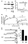
NMDARs are activated by [glu]o during ED. (A1) Single trace depicting that ED-induced outward current in CA3 pyramidal cells is decreased by dAPV. Downward deflections represent responses to −10 mV voltage steps. (Inset) The current–voltage relationship of dAPV-sensitive current displays a negative slope at hyperpolarized potentials, characteristic of NMDAR currents. (A2) Pooled data; n = 17. (A3) dAPV reduces the current to 30 ± 5% of its initial value. (A4) Under Ctl conditions, dAPV blocks outward currents induced by glutamate pulses (400 μM in pipette; 150 ms) without affecting baseline holding current. (B1) Single trace depicting that responses to NMDA pulses decrease during ED. (B2) Pooled data; n = 14. (C1) An increase in ambient [glu]o is not sufficient to occlude NMDAR-mediated responses. Time course of the effect of TBOA on baseline holding current and NMDAR-mediated responses to NMPA and glutamate puffs. (C2) Time course of the effect of TBOA on NMDA and glutamate puffs after subtraction of baseline holding current. (D1) Depression of NMDAR-mediated EPSCs. (D2) Time-course graph. Stimulus frequency, 0.1 Hz; bin size, six stimuli; n = 4. dAPV concentration is 100 μM in this and in subsequent figures.
Figure 2
Figure 2
ED increases AP-independent vesicular release of glutamate. (A1) Sample traces of AMPA receptor-mediated miniature EPSCs at −60 mV, in the presence of dAPV and picrotoxin at two different time resolutions. (A2) Time-course plot of the increase in mEPSC frequency during ED. (A3) Cumulative plot for interevent intervals. (Inset) Expanded time scale; n = 5. (B1) Inhibition of vesicular release with TeNT limits the increase in [glu]o during ED. (B2) Summary histogram of normalized values of the dAPV-sensitive current after 9 min of ED for Ctl and TeNT-treated cultures; n = 9.
Figure 3
Figure 3
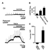
Increased vesicular release is not sufficient to account for increased [glu]o. (A) (Top) Increasing mEPSC frequency with sucrose under Ctl conditions induces negligible outward current. (Bottom) After blockade of glutamate uptake with TBOA, sucrose induces a large NMDAR-mediated outward current (different cell from above). (B) The frequency of mEPSCs is not significantly different after 2 min treatment with sucrose or 6 min of ED. Values are normalized to mEPSC frequency during Ctl; n = 8. (C) Pooled data for the amplitude of NMDAR-mediated currents induced by sucrose alone and by sucrose in presence of TBOA; n = 6.
Figure 4
Figure 4
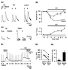
ED reduces net glutamate uptake. (A1) Brief alternating applications of glutamate and NMDA were used to evaluate the net effect of glutamate transporter function on glutamate responses. In the sample traces, both responses decrease in amplitude, but the NMDA response is decreased to a greater extent than is the glutamate response. (Right) Both traces were scaled to the NMDA response amplitude under Ctl conditions. (A2) Pooled data. (Upper) Glutamate responses decrease less than NMDA responses do. (Lower) The glutamate/NMDA response ratio reveals the time course of the rundown in glutamate transporter function. Applications were alternated every 15 s. Bin size, four applications; n = 8. The arrowhead indicates the time when sample traces in A1 were recorded. (A3) Inhibition of glutamate transporters with TBOA (250 μM) increases the glutamate/NMDA response ratio. Note that NMDA responses are unaffected. Traces show averages of three consecutive responses. (B1) TBOA increases [glu]o under Ctl conditions, but its effect is occluded during ED. In the sample trace, upward deflections are responses to 200-ms NMDA applications. (B2) Illustration of data from individual cells. The open circles denote the cell in A1; the closed circles denote the other four cells tested. (B3) Pooled values; n = 5. Note that TBOA responses are depressed by a factor of 8 as compared with NMDA responses.
Figure 5
Figure 5
ED promotes reverse glutamate transport. (A) Glutamate heteroexchange increases during ED. Sample trace showing that brief (300 ms) tPDC applications induce progressively more release of glutamate during ED. (A2) Pooled data; n = 7. (B) Glutamate efflux can occur during reverse transport or during heteroexchange. Pulses of tPDC will shift the transporter into the “glu/3Na+/H+ inside” state. The glutamate binding site is then returned to the outside through transport of either K+ or glutamate. During ED, the latter pathway (common to heteroexchange and reverse transport) is favored. See Results for details. This figure is adapted from refs. and .

"V体育官网入口" References

    1. Choi D W, Rothman S M. Annu Rev Neurosci. 1990;13:171–182. - PubMed
    1. Martin R L, Lloyd H G, Cowan A I. Trends Neurosci. 1994;17:251–257. - PubMed
    1. Obrenovitch T P, Urenjak J. Prog Neurobiol. 1997;51:39–87. - PubMed (VSports app下载)
    1. Drejer J, Benveniste H, Diemer N H, Schousboe A. J Neurochem. 1985;45:145–151. - PubMed
    1. Bosley T M, Woodhams P L, Gordon R D, Balazs R. J Neurochem. 1983;40:189–201. - PubMed

"V体育官网入口" Publication types

VSports - MeSH terms

Substances