<code lang="wl1MLOOg"></code> Skip to main page content (V体育ios版)
U.S. flag

An official website of the United States government

Dot gov

The . gov means it’s official. Federal government websites often end in VSports app下载. gov or . mil. Before sharing sensitive information, make sure you’re on a federal government site. .

Https

The site is secure. The https:// ensures that you are connecting to the official website and that any information you provide is encrypted and transmitted securely V体育官网. .

. 2000 May;20(9):3157-67.
doi: 10.1128/MCB.20.9.3157-3167.2000.

"VSports在线直播" STRAP and Smad7 synergize in the inhibition of transforming growth factor beta signaling

Affiliations

V体育官网入口 - STRAP and Smad7 synergize in the inhibition of transforming growth factor beta signaling

P K Datta et al. Mol Cell Biol. 2000 May.

Abstract (V体育2025版)

Smad proteins play a key role in the intracellular signaling of the transforming growth factor beta (TGF-beta) superfamily of extracellular polypeptides that initiate signaling from the cell surface through serine/threonine kinase receptors. A subclass of Smad proteins, including Smad6 and Smad7, has been shown to function as intracellular antagonists of TGF-beta family signaling. We have previously reported the identification of a WD40 repeat protein, STRAP, that associates with both type I and type II TGF-beta receptors and that is involved in TGF-beta signaling VSports手机版. Here we demonstrate that STRAP synergizes specifically with Smad7, but not with Smad6, in the inhibition of TGF-beta-induced transcriptional responses. STRAP does not show cooperation with a C-terminal deletion mutant of Smad7 that does not bind with the receptor and consequently has no inhibitory activity. STRAP associates stably with Smad7, but not with the Smad7 mutant. STRAP recruits Smad7 to the activated type I receptor and forms a complex. Moreover, STRAP stabilizes the association between Smad7 and the activated receptor, thus assisting Smad7 in preventing Smad2 and Smad3 access to the receptor. STRAP interacts with Smad2 and Smad3 but does not cooperate functionally with these Smads to transactivate TGF-beta-dependent transcription. The C terminus of STRAP is required for its phosphorylation in vivo, which is dependent on the TGF-beta receptor kinases. Thus, we describe a mechanism to explain how STRAP and Smad7 function synergistically to block TGF-beta-induced transcriptional activation. .


Figures
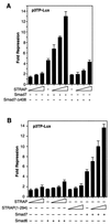
FIG. 1
FIG. 1
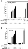
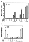
Synergy between STRAP and Smad7 in the inhibition of p3TP promoter activity in response to TGF-β. (A) STRAP synergizes with Smad7 but not with mutant Smad7-Δ408. Mv1Lu cells were transiently transfected with p3TP-Lux (0.3 μg), the β-galactosidase reporter (30 ng), and TβR-I(TD) (0.43 μg) and with Smad7 constructs (0.3 μg) and increasing amounts of STRAP (0.2, 0.5, and 1 μg) as indicated. In each experiment equal amounts of total DNA were transfected. Luciferase activity was normalized to β-galactosidase activity. The mean of triplicate luciferase values from the TGF-β-treated control was considered 100%, and this was then divided by values for three replicates of each point to get the fold repressions. The means of these fold repressions ± standard deviations are plotted. These experiments were performed four times in triplicate with similar results. (B) STRAP does not synergize with Smad6, but the STRAP(1-294) mutant shows synergy with Smad7. Mv1Lu cells were transfected as described above with Smad7 or Smad6 (0.52 μg) and increasing amounts of STRAP or STRAP(1-294). Luciferase assays were performed as described for panel A.
FIG. 2
FIG. 2
Functional synergy between STRAP and Smad7. (A) Synergistic inhibition of (CAGA)9 MLP-Luc reporter activity in response to TGF-β. HepG2 cells were transfected with a (CAGA)9 MLP-Luc reporter (0.3 μg) containing nine copies of Smad3/Smad4 binding sites, Smad7 constructs, increasing amounts of STRAP, and increasing amounts of STRAP(1-294) (0.5 and 1 μg). TGF-β signaling was initiated by expression of TβR-I(T204D). Luciferase assays were performed as described for Fig. 1A. (B) STRAP and Smad7 synergistically block an immediate-early response to TGF-β. HepG2 cells were cotransfected with pAR3-lux (0.3 μg), FAST2 (15 ng), Smad7 constructs, STRAP(1-294) (1 μg), and increasing amounts of STRAP as indicated. Cells were treated with or without TGF-β (100 pM) for 20 h prior to lysis and then analyzed for luciferase activity. (C) Synergistic inhibition of TGF-β-induced PAI-1 promoter activity by STRAP and Smad7. HepG2 cells were transiently transfected with pGLuc 884 reporter (0.25 μg) (9), HA-tagged Smad7 constructs, and increasing amounts of STRAP. TGF-β signaling was initiated either by treatment of the cells with 100 pM TGF-β (left) or by coexpression of TβR-I(TD) (right). Luciferase assays were performed as described for Fig. 1A. Expression of Smad7 proteins were confirmed by direct immunoblotting of total cell lysates, made for luciferase assays from cells transfected with either vector or coding sequences for Smad7 or the Smad7-Δ408 construct, with anti-HA antibodies.
FIG. 2
FIG. 2
Functional synergy between STRAP and Smad7. (A) Synergistic inhibition of (CAGA)9 MLP-Luc reporter activity in response to TGF-β. HepG2 cells were transfected with a (CAGA)9 MLP-Luc reporter (0.3 μg) containing nine copies of Smad3/Smad4 binding sites, Smad7 constructs, increasing amounts of STRAP, and increasing amounts of STRAP(1-294) (0.5 and 1 μg). TGF-β signaling was initiated by expression of TβR-I(T204D). Luciferase assays were performed as described for Fig. 1A. (B) STRAP and Smad7 synergistically block an immediate-early response to TGF-β. HepG2 cells were cotransfected with pAR3-lux (0.3 μg), FAST2 (15 ng), Smad7 constructs, STRAP(1-294) (1 μg), and increasing amounts of STRAP as indicated. Cells were treated with or without TGF-β (100 pM) for 20 h prior to lysis and then analyzed for luciferase activity. (C) Synergistic inhibition of TGF-β-induced PAI-1 promoter activity by STRAP and Smad7. HepG2 cells were transiently transfected with pGLuc 884 reporter (0.25 μg) (9), HA-tagged Smad7 constructs, and increasing amounts of STRAP. TGF-β signaling was initiated either by treatment of the cells with 100 pM TGF-β (left) or by coexpression of TβR-I(TD) (right). Luciferase assays were performed as described for Fig. 1A. Expression of Smad7 proteins were confirmed by direct immunoblotting of total cell lysates, made for luciferase assays from cells transfected with either vector or coding sequences for Smad7 or the Smad7-Δ408 construct, with anti-HA antibodies.
FIG. 3
FIG. 3
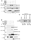
Association of STRAP with Smad7, but not with Smad7-Δ408, and oligomerization of STRAP. (A) Interaction of STRAP with Smad6 and Smad7 in mammalian cells. COS-1 cells were transfected with HA-tagged STRAP either alone or together with the indicated Flag-tagged Smad constructs, including Smad1(AAVA), Smad6, and Smad7. Cell lysates were subjected to an anti-Flag immunoprecipitation (IP), and coprecipitating STRAP was detected by immunoblotting (Blot) with anti-HA antibodies (top section). In the middle section, total lysates were immunoprecipitated using anti-HA antibodies and then immunoblotted with anti-Flag antibodies. To confirm expression of Smads, aliquots of total cell lysates were immunoblotted with anti-Flag antibodies (bottom section). Ig, immunoglobulin. (B) STRAP(1-294) interacts with Smad7, and Smad7-Δ408 does not interact with STRAP. COS-1 cells were transiently transfected with the indicated combinations of Flag-tagged STRAP constructs and HA-tagged Smad7 constructs. Cell lysates were immunoprecipitated with an anti-Flag antibody, and the immunoprecipitates were analyzed by anti-HA antibody immunoblotting (top section). In the second section from the top, cell lysates were subjected to immunoprecipitation with an anti-HA antibody and the precipitates were analyzed with an anti-Flag antibody. Expression of the proteins was confirmed by the direct immunoblotting of the total cell lysates (bottom two sections). (C) Homo-oligomerization of STRAP. Cells were transfected with STRAP-HA alone or together with STRAP-Flag or TβR-II-Flag (serves as a positive control) as indicated. Cell lysates were subjected to immunoprecipitation with a Flag antibody, and coprecipitated proteins were detected by immunoblotting with an HA antibody (lanes 1 to 4). Reciprocal experiments were also performed (lanes 5 and 6).
FIG. 4
FIG. 4
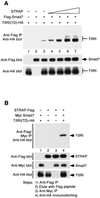
STRAP stabilizes the association between Smad7 and activated TβR-I and forms a complex with Smad7 and TβR-I(TD). (A) STRAP stabilizes Smad7–TβR-I(TD) complexes. COS-1 cells were transiently transfected with plasmids encoding Flag-Smad7 (0.4 μg), TβR-I(TD)-HA (0.6 μg), and STRAP (in increasing amounts of 0.2, 0.4, 1, and 2 μg). Cell lysates were subjected to immunoprecipitation (IP) with an anti-Flag antibody, and the presence of TβR-I(TD) in the immunoprecipitates was detected by immunoblotting with an anti-HA antibody (top). To confirm equivalent expression of Smad7 and TβR-I(TD), aliquots of total cell lysates were immunoblotted with an anti-Flag antibody (middle) and an anti-HA antibody (bottom). (B) STRAP is present in a complex with Smad7 and TβR-I(TD). Cells were transfected with indicated combinations of STRAP-Flag, Myc-Smad7, and TβR-I(TD)-HA. Cell lysates were immunoprecipitated with an anti-Flag antibody, proteins were eluted with a Flag peptide, and the eluate was reprecipitated by an anti-Myc antibody followed by anti-HA antibody immunoblotting (top). Expression of the proteins was monitored by immunoblotting.
FIG. 5
FIG. 5
Interaction between STRAP and Smad7 is stronger than that between STRAP and Smad2 or between STRAP and Smad3. (A) STRAP interacts with Smad2 and Smad3. COS-1 cells were transiently transfected with combinations of HA-tagged STRAP and Flag-tagged Smad2 or Smad3 as indicated. Cell lysates were subjected to immunoprecipitation (IP) with anti-Flag antibodies and then immunoblotted (Blot) using anti-HA antibodies (top section). In the second section from the top, total lysates were immunoprecipitated using anti-HA antibodies and immunoblotted with anti-Flag antibodies. To confirm the expression of the proteins, aliquots of total cell lysates were immunoblotted with anti-Flag antibodies (third section from the top) and anti-HA antibodies (bottom section). IgH, immunoglobulin H. (B) Relative strengths of binding of STRAP with Smad2, Smad3, and Smad7. COS-1 cells were transfected with a constant amount of the STRAP-HA construct together with increasing amounts of Flag-Smad7 (top), Flag-Smad3 (middle), or Flag-Smad2 (bottom). Cell lysates were subjected to immunoprecipitation with anti-HA antibodies, and the precipitates were analyzed by blotting with anti-Flag antibodies (all three sections). Expression of Smad proteins was monitored by analyzing aliquots of total cell lysate by immunoblotting with anti-Flag antibodies (all sections). Smad7, Smad3, and Smad2 were expressed in equivalent levels in lanes 2 to 5. In lane 6 (middle and bottom), Smad3 and Smad2 were expressed at levels about twofold higher than the corresponding levels of these proteins in lane 5. Expression of STRAP-HA was determined by immunoblotting total cell lysates using anti-HA antibodies. The migration of each protein is indicated on the right by an arrow, and the arrowhead points to the expected position of Smad2 in the bottom section.
FIG. 6
FIG. 6
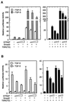
Inhibition of TGF-β-induced and Smad3- or Smad2-dependent transcription by STRAP. (A) Smad3-dependent transcriptional activation of the p3TP promoter is not enhanced by STRAP. HepG2 cells were transiently transfected with p3TP-Lux, Smad3, and increasing amounts of STRAP as indicated. Left, cells were treated with or without TGF-β (100 pM) for 20 h prior to lysis and then analyzed for luciferase activity. Right, TGF-β signaling was initiated by expression of TβR-I(TD). Luciferase activity was normalized to β-galactosidase activity and expressed as the mean ± standard deviation of triplicate measurements from a representative experiment. These experiments were performed four times in triplicate with similar results. (B) STRAP does not cooperate with Smad2 in its transactivation function. This experiment was same as that in panel A except that Smad2 was expressed instead of Smad3.
FIG. 7
FIG. 7
Smad3, and not Smad2, strongly reverses the inhibition of TGF-β-induced transcription by either Smad7 alone or STRAP and Smad7 together. (A) Reversing the synergistic inhibition of TGF-β-mediated transcription by Smad3. HepG2 cells were transfected with p3TP-Lux and with coding sequences for Smad7, STRAP, and increasing amounts of Smad3 as indicated and were treated with or without TGF-β (100 pM) for 20 h. The relative luciferase activity in cell lysates was measured. Luciferase activity was normalized to β-galactosidase activity and expressed as the mean ± standard deviation of triplicate measurements from a representative experiment. These experiments were performed four times in triplicate with similar results. (B) Smad2 shows a weak effect on reversing the inhibition of the promoter activity by STRAP and Smad7. The experiment was performed as described for panel A except that Smad2 was transfected in two doses instead of Smad3.
FIG. 8
FIG. 8
The C terminus of STRAP is required for its TGF-β receptor-dependent phosphorylation. (A) Phosphorylation of STRAP through its C terminus. COS-1 cells were transiently transfected with STRAP-Flag or STRAP(1-294)-Flag in combination with wild-type (wt) or kinase-defective HA-tagged TβR-I and/or hexahistidine-tagged TβR-II as indicated. Cells were metabolically labeled with [32P]orthophosphate, and equal amounts of extracts were immunoprecipitated with an anti-Flag antibody. Phosphorylated STRAP was detected by SDS-PAGE and autoradiography (top). Equivalent levels of expression of STRAP-Flag and STRAP(1-294)-Flag proteins was confirmed by immunoblotting total cell lysates (middle). Phosphate incorporated into STRAP is plotted in relative units (bottom). The result is representative of five independent experiments. (B) Confirmation of the phosphorylated band as STRAP. The immunoprecipitate from lane 6 of panel A (lane 1) was boiled with Laemmli sample buffer to disrupt the complex and then was subjected to a second immunoprecipitation with anti-Flag antibody (lane 2).

References

    1. Abdollah S, Macias-Silva M, Tsukazaki T, Hayashi H, Attisano L, Wrana J L. TβRI phosphorylation of Smad2 on serine 465 and 467 is required for Smad2/Smad4 complex formation and signaling. J Biol Chem. 1997;272:27678–27685. - VSports最新版本 - PubMed
    1. Attisano L, Wrana J L, López-Casillas F, Massagué J. TGF-β receptors and actions. Biochim Biophys Acta. 1994;1222:71–80. - PubMed
    1. Barinaga M. New clues to how proteins link up to run the cell. Science. 1999;283:1247–1249. - PubMed
    1. Caŕcamo J, Weis F M B, Ventura F, Wieser R, Wrana J L, Attisano L, Massagué J. Type I receptors specify growth inhibitory and transcriptional responses to TGF-β and activin. Mol Cell Biol. 1994;14:3810–3821. - PMC - PubMed
    1. Chen R-H, Derynck R. Homomeric interactions between type II transforming growth factor-β receptors. J Biol Chem. 1994;269:22868–22874. - PubMed

Publication types

MeSH terms

"VSports最新版本" Substances

LinkOut - more resources