Skip to main page content
U.S. flag

An official website of the United States government

Dot gov

The . gov means it’s official. Federal government websites often end in VSports app下载. gov or . mil. Before sharing sensitive information, make sure you’re on a federal government site. .

Https

The site is secure. The https:// ensures that you are connecting to the official website and that any information you provide is encrypted and transmitted securely V体育官网. .

. 2000 Mar 15;19(6):1301-11.
doi: 10.1093/emboj/19.6.1301.

Molecular determinants that mediate selective activation of p38 MAP kinase isoforms

Affiliations

Molecular determinants that mediate selective activation of p38 MAP kinase isoforms

H Enslen et al. EMBO J. .

Abstract

The p38 mitogen-activated protein kinase (MAPK) group is represented by four isoforms in mammals (p38alpha, p38beta2, p38gamma and p38delta). These p38 MAPK isoforms appear to mediate distinct functions in vivo due, in part, to differences in substrate phosphorylation by individual p38 MAPKs and also to selective activation by MAPK kinases (MAPKKs). Here we report the identification of two factors that contribute to the specificity of p38 MAPK activation. One mechanism of specificity is the selective formation of functional complexes between MAPKK and different p38 MAPKs. The formation of these complexes requires the presence of a MAPK docking site in the N-terminus of the MAPKK VSports手机版. The second mechanism that confers signaling specificity is the selective recognition of the activation loop (T-loop) of p38 MAPK isoforms. Together, these processes provide a mechanism that enables the selective activation of p38 MAPK in response to activated MAPKK. .


Figures

None
Fig. 1. Identification of a domain required for activation of p38β2 by MAP kinase kinases. (A) Schematic representation of MKK3, MKK3b, MKK6 and chimeras. MKK6 is shown in white, MKK3 in black and the N–terminal extension of MKK3b is gray. In the chimeras, domains from MKK3 are shown in black and domains from MKK6 in white. (B and C) Epitope-tagged p38α (B) or p38β2 (C) were immunoprecipitated from COS7 cells co-transfected with an empty vector (Control) or activated MAPKK. The activated MAPKKs were constructed by replacing the two sites of activating phosphorylation with glutamic acid residues. Immune complex kinase assays were performed to measure p38 MAP kinase activity using ATF2 as the substrate. The expression of MAPKK and p38 was examined by immunoblot analysis (lower panel). The rate of ATF2 phosphorylation was quantitated by PhosphorImager analysis and is presented as relative protein kinase activity.
None
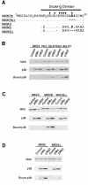
Fig. 2. Binding of p38α and p38β2 to MAP kinase kinases. (A) Primary sequences of the N–terminal domain of MKK3, MKK3b, MKK6 and deletion mutants (Δ) are aligned. Residues that are identical to MKK3b are indicated with a dot (.). The residues of MKK3b (LRI) and MKK6 (LKI) deleted in MKK3bΔ and MKK6Δ are indicated in bold. The deleted residues are indicated with a dash (–). Basic residues are indicated by asterisks. (B) Activated GST-tagged MKK3, K6(1–18)–K3 or K6(1–82)–K3 were co-transfected with an empty vector (Control), Flag-tagged p38α or Flag-tagged p38β2 in COS7 cells. The activated MKKs were constructed by replacing the two sites of activating phosphorylation with glutamic acid residues. Protein expression was monitored by immunoblot analysis of cell extracts. The GST–MKK fusion proteins were isolated from the cell extracts by incubation with glutathione–Sepharose. The co-precipitation of p38α and p38β2 with the MKK was examined by immunoblot analysis with an antibody to the Flag epitope. (C and D) The interaction of Flag-tagged p38α and p38β2 with GST-tagged activated MKK3, MKK3b and MKK3bΔ (C) or MKK6 and MKK6Δ (D) co-expressed in COS7 cells was examined using the methods described in (B). The activated MKKs were constructed by replacing the two sites of activating phosphorylation with glutamic acid residues.
None
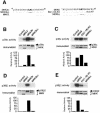
Fig. 3. Regulation of p38α and p38β2 by MAP kinase kinases. (A) The primary sequences of the N–terminal region of MKK3, MKK3b and MKK3bΔ (left panel) and of MKK6, K6(1–18)–K3 and MKK6Δ (right panel) are aligned. (B–E) Flag-tagged p38α (B and C) or p38β2 (D and E) were co-transfected together with activated MKK3, MKK3b and MKK3bΔ (B and D) or activated MKK6, K6(1–18)–K3 and MKK6Δ (C and E) in COS7 cells. The activated MKKs were constructed by replacing the two sites of activating phosphorylation with glutamic acid residues. The expression of p38 and MKK was examined by immunoblot analysis of cell lysates. The protein kinase activity of p38α and p38β2 was measured in immune complex kinase assays using ATF2 as the substrate. The phosphorylated ATF2 was detected after SDS–PAGE by auto- radiography and was quantitated by PhosphorImager analysis. The p38 activity is presented as relative protein kinase activity.
None
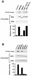
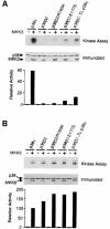
Fig. 4. Regulation of endogenous p38α activity by MAP kinase kinases. Activated epitope-tagged MKK3b, MKK3, K6(1–18)–K3 and K6(1–82)–K3 (A) or MKK3b, MKK3bΔ, MKK6, MKK6Δ and MKK3 (B) were expressed in COS7 cells. The activated MKKs were constructed by replacing the two sites of activating phosphorylation with glutamic acid residues. The epitope-tagged MKK expression and endogenous p38α were examined by immunoblot analysis of cell lysates. The activity of the endogenous p38α was measured in immune complex kinase assays using ATF2 as the substrate. The phosphorylated ATF2 was detected after SDS–PAGE by auto- radiography and was quantitated by PhosphorImager analysis. The p38 activity is presented as relative protein kinase activity.
None
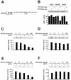
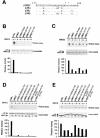
Fig. 5. Inhibition of p38β2 activity by peptide competition. (A) The primary sequence of synthetic peptides corresponding to the native (wt-pep) or mutated (gly-pep) N–terminal region of MKK3b is shown. The mutated peptide was prepared by replacing the residues of MKK3b (LRI), indicated in bold, with glycine. (B) Purified bacterially expressed GST–p38α was bound to glutathione–Sepharose and incubated with 100 μM wild-type or mutated MKK3b peptide. Purified activated MKK3, MKK3b or MKK6 were incubated with the immobilized GST–p38α in kinase buffer with ATP for 20 min. The GST–p38α was washed with kinase buffer and the p38α activity was measured using ATF2 and [γ-32P]ATP as the substrates. The phosphorylated ATF2 was detected after SDS–PAGE by auto- radiography and was quantitated by PhosphorImager analysis. The p38α activity is presented as relative protein kinase activity. (CF) Purified bacterially expressed GST–p38β2 was bound to glutathione–Sepharose and incubated with increasing concentrations of the wild-type (C and E) or mutated (D and F) MKK3b peptide. Purified activated MKK3b (C and D) or MKK6 (E and F) were incubated with the immobilized GST–p38β2 in kinase buffer with ATP for 20 min. The GST–p38β2 activity was measured as described in (B).
None
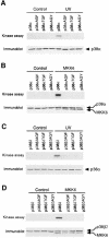
Fig. 6. Dual phosphorylation on threonine and tyrosine is required for p38 MAPK activation. Epitope-tagged wild-type and mutated p38α (A and B) or p38β2 (C and D) were expressed in COS7 cells. The mutated p38 contained point mutations in the Thr-Gly-Tyr (TGY) dual phosphorylation motif (replacement of threonine and tyrosine with alanine and phenylalanine, respectively). The effect of treatment with 80 J/m2 UV (A and C) or co-expression with activated MKK6 (B and D) was examined. Immune complex kinase assays were performed to measure p38 activity using ATF2 as the substrate. The expression of p38 and activated MKK6 was examined by immunoblot analysis (lower panel).
None
Fig. 7. Phosphoamino acid analysis of p38α and p38β2 phosphorylated by MAP kinase kinases. Activated epitope-tagged MKKs (described in Figure 1A) were expressed in COS7 cells and isolated by immunoprecipitation. Immune complex kinase assays were performed using [γ-32P]ATP and purified bacterially expressed GST–p38α or GST–p38β2 as the substrates. The phosphorylated p38 was examined by phosphoamino acid analysis. The figure shows an autoradiograph of the phosphoamino acids separated by thin layer electrophoresis. The relative phosphorylation on serine, threonine and tyrosine was examined by PhosphorImager analysis.
None
Fig. 8. Regulation of chimeric p38 MAPK by MKK3 and MKK6. (A) Schematic representation of p38α, p38β2 and chimeric protein kinases. Regions of p38α are shown in black and regions of p38β2 in white. The p38β2-TL-p38α was constructed by replacing residues within the T–loop of p38β2 with those derived from p38α. (B and C) Epitope-tagged p38α, p38β2 or p38 chimeras were expressed in COS7 cells together with an empty vector (–) and activated (+) MKK3 (B) or MKK6 (C) The expression of MKK and p38 was examined by immunoblot analysis. The p38 activity was measured in immune complex kinase assays with ATF2 as the substrate. The rate of phosphorylation was quantitated by PhosphorImager analysis and is presented as relative p38 activity.
None
Fig. 9. Effect of p38α T–loop mutations on p38β2 activation. (A) Comparison of the sequences of p38α, p38β2, p38γ and p38δ within the dual phosphorylation domain (T–loop). Residues in p38 isoforms that are identical to p38β2 are indicated with a dot (.). The sites of activating phosphorylation (threonine and tyrosine) are indicated with asterisks. (BE) Flag-tagged p38α, p38β2 or T–loop mutants of p38β2 were expressed in COS7 cells and p38 activity was measured in immune complex kinase assays using ATF2 as the substrate. The effect of co-expression together with an empty vector (–) and activated (+) MKK3 (B and D) or activated MKK6 (C and E) was examined. The expression of MKK3, MKK6 and p38 was monitored by immunoblot analysis. The rate of phosphorylation was quantitated by PhosphorImager analysis and is presented as relative p38 activity.
None
Fig. 10. Effect of p38γ T–loop mutations on p38β2 activation. Flag-tagged p38γ, p38β2 or T–loop mutants of p38β2 were expressed in COS7 cells and p38 activity was measured in immune complex kinase assays using ATF2 as the substrate. The effect of co-expression together with an empty vector (–) and activated (+) MKK3 (A) or activated MKK6 (B) was examined. The expression of MKK3, MKK6 and p38 was monitored by immunoblot analysis. The rate of phosphorylation was quantitated by PhosphorImager analysis and is presented as relative p38 activity.
None
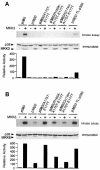
Fig. 11. Effect of p38δ T–loop mutations on p38β2 activation. Flag-tagged p38δ, p38β2 or T–loop mutants of p38β2 were expressed in COS7 cells, and p38 activity was measured in immune complex kinase assays using ATF2 as the substrate. The effect of co-expression together with an empty vector (–) and activated (+) MKK3 (A) or activated MKK6 (B) was examined. The expression of MKK3, MKK6 and p38 was monitored by immunoblot analysis. The rate of phosphorylation was quantitated by PhosphorImager analysis and is presented as relative p38 activity.

References (VSports app下载)

    1. Bardwell L. and Thorner, J. (1996) A conserved motif at the amino termini of MEKs might mediate high-affinity interaction with the cognate MAPKs. Trends Biochem. Sci., 21, 373–374. - PubMed
    1. Bardwell L., Cook, J.G., Chang, E.C., Cairns, B.R. and Thorner, J. (1996) Signaling in the yeast pheromone response pathway: specific and high-affinity interaction of the mitogen-activated protein (MAP) kinases Kss1 and Fus3 with the upstream MAP kinase kinase Ste7. Mol. Cell. Biol., 16, 3637–3650. - VSports注册入口 - PMC - PubMed
    1. Brunet A. and Pouyssegur, J. (1996) Identification of MAP kinase domains by redirecting stress signals into growth factor responses. Science, 272, 1652–1655. - PubMed
    1. Camps M., Nichols, A., Gillieron, C., Antonsson, B., Muda, M., Chabert, C., Boschert, U. and Arkinstall, S. (1998) Catalytic activation of the phosphatase MKP-3 by ERK2 mitogen-activated protein kinase. Science, 280, 1262–1265. - VSports注册入口 - PubMed
    1. Chow C.W., Rincon, M., Cavanagh, J., Dickens, M. and Davis, R.J. (1997) Nuclear accumulation of NFAT4 opposed by the JNK signal transduction pathway. Science, 278, 1638–1641. - PubMed

Publication types

MeSH terms

Substances

LinkOut - more resources