<u dropzone="qJSOjVp"></u> Skip to main page content
U.S. flag

An official website of the United States government

Dot gov

The . gov means it’s official. Federal government websites often end in . gov or . mil. Before sharing sensitive information, make sure you’re on a federal government site VSports app下载. .

Https

The site is secure. The https:// ensures that you are connecting to the official website and that any information you provide is encrypted and transmitted securely. V体育官网.

. 1999 May 25;96(11):6211-6.
doi: 10.1073/pnas.96.11.6211.

CMS: an adapter molecule involved in cytoskeletal rearrangements

Affiliations

CMS: an adapter molecule involved in cytoskeletal rearrangements

K H Kirsch et al. Proc Natl Acad Sci U S A. .

Abstract

Cas ligand with multiple Src homology (SH) 3 domains (CMS) is an ubiquitously expressed signal transduction molecule that interacts with the focal adhesion protein p130(Cas). CMS contains three SH3 in its NH2 terminus and proline-rich sequences in its center region. The latter sequences mediate the binding to the SH3 domains of p130(Cas), Src-family kinases, p85 subunit of phosphatidylinositol 3-kinase, and Grb2. The COOH-terminal region contains putative actin binding sites and a coiled-coil domain that mediates homodimerization of CMS. CMS is a cytoplasmic protein that colocalizes with F-actin and p130(Cas) to membrane ruffles and leading edges of cells. Ectopic expression of CMS in COS-7 cells resulted in alteration in arrangement of the actin cytoskeleton. We observed a diffuse distribution of actin in small dots and less actin fiber formation. Altogether, these features suggest that CMS functions as a scaffolding molecule with a specialized role in regulation of the actin cytoskeleton. VSports手机版.


Figures
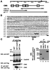
Figure 1
Figure 1
Schematic representation of the human CMS and protein sequence. (A) Alignment of human cDNA clones isolated in the yeast two-hybrid screen, 5′ rapid amplification of complementary DNA ends (RACE) and λgt11 cDNA library screen with respect to the full-length CMS cDNA. PR, proline-rich region. (B) Predicted protein sequence of CMS. Sequence numbers are shown on the left. The SH3 domains are underlined. Proline-rich sequences are marked in bold, the CC domain is marked in italics, and the putative actin binding sites are bold underlined. IP, immunoprecipitation. (C) In vitro interaction of p130Cas and CMS. 293T cells were transiently transfected with the flag-tagged CMS yeast TH clone (CMS-TH) together with the CasSH3 domain or full-length Cas (both GST tagged) or the vector alone (GST control). Cell lysates were immunoprecipitated with anti-flag antibody or glutathione Sepharose beads, and precipitates were subjected to SDS/PAGE and probed with anti-GST antibody and anti-flag antibody, respectively. The upper blot was striped and reprobed with anti-flag antibody. (D) Direct interaction of CMS with p130Cas. Five hundred micrograms of protein from 293T cells expressing the various CMS peptides or C3G used as a positive control was immunoprecipitated with the antibodies indicated above. Blots were probed for binding with 32P-labeled GST-p130CasSH3 domain.
Figure 2
Figure 2
Tissue distribution of CMS mRNA. (A) Northern blot analysis of poly(A)+ RNA from adult human tissues by using the 32P-labeled CMS cDNA clone isolated in the two-hybrid screen. Hybridization analysis of the same Northern blot by using a 32P-labeled β-actin cDNA probe (Lower). (B) Relative expression levels of CMS in different human tissues and developmental stages. Adult tissue: A1, whole brain; A2, amygdala; A3, caudate nucleus; A4, cerebellum; A5, cerebral cortex; A6, frontal lobe; A7, hippocampus; A8, medulla oblongata; B1, occipital lobe; B2, putamen; B3, substantia nigra; B4, temporal lobe; B5, thalamus; B6, nucleus accumbeus; B7, spinal cord; C1, heart; C2, aorta; C3, skeletal muscle; C4, colon; C5, bladder; C6, uterus; C7, prostate; C8, stomach; D1, testis; D2, ovary, D3, pancreas, D4, pituitary gland; D5, adrenal gland; D6, thyroid gland; D7, salivary gland; D8, mammary gland; E1, kidney; E2, liver; E3, small intestine; E4, spleen; E5, thymus; E6, peripheral leukocyte; E7, lymph node; E8, bone marrow; F1, appendix; F2, lung; F3, trachea; F4, placenta. Fetal tissue: G1, brain; G2, heart; G3, kidney; G4, liver; G5, spleen; G6, thymus; G7, lung. Controls (100 ng each): H1, yeast total RNA; H2, yeast tRNA; H3, Escherichia coli rRNA; H4, E. coli DNA; H5, poly(rA); H6, human C0t 1 DNA; H7, human DNA; H8, human DNA (500 ng).
Figure 3
Figure 3
CMS is localized in the cytoplasm and colocalizes with actin and p130Cas to membrane ruffles. (A and B) COS-7 cells grown in medium containing 10% FBS, plated on poly-d-lysine coverslips, were transfected with the flag-tagged CMS constructs CMS, CMS/TH, and CMS/3SH3 as indicated. Cells were fixed and stained with rhodamine-labeled phalloidin (lane 1) or with anti-flag antibody (lanes 3 and 4). Merged images are shown in lane 2. Actin clustering adjacent to the nucleus (B, lane 1 marked with an arrow). Vesicles in CMS cells are indicated by arrows (B, lane 3). (C and D) Membrane ruffle induction in COS-7 cells transfected with flag-tagged CMS or GST-tagged p130Cas. Serum-starved cells were treated with 10 nM phorbol 12-myristate 13-acetate (PMA) for 10 min, fixed, and subjected to immunofluorescence analysis by using the anti-flag or p130Cas antibody (lane 3). Actin filaments were visualized by staining with rhodamine-labeled phalloidin (lane 1). The cells were analyzed by confocal microscopy (×250 and ×630). Yellow-orange color in merged image shows colocalization of CMS and p130Cas with actin (lane 2, marked with arrows). (Bar, 20 μm.)
Figure 4
Figure 4
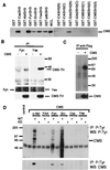
Analysis of SH3-containing proteins for their interaction with CMS. (A) GST-fusion peptides coupled to glutathione Sepharose beads were incubated with lysates of 293T cells expressing flag-tagged CMS. Precipitates were analyzed by SDS/PAGE and WB with anti-flag antibody. (B) In vivo interaction of CMS with the tyrosine kinases Fyn and Yes. Lysates of 293T cells expressing flag-tagged CMS-TH or control vector were incubated with anti-Fyn and anti-Yes antibody coupled to protein A/G agarose beads. Immunoprecipitates (IP) were analyzed in SDS/PAGE and WB with anti-flag antibody. (C) Kinase activity is associated with CMS. Lysates of 293T cells expressing flag-tagged CMS or vector control were incubated with anti-flag antibody coupled to protein A/G agarose, and immunoprecipitates (IP) were subjected to a kinase assay. (D) Tyrosine phosphorylation of CMS. 293T cell lysates expressing flag-tagged CMS together with the indicated tyrosine kinases [wild type (WT) or kinase dead (KD)] were immunoprecipitated (IP) with antiphoshotyrosine antibody (4G10) and analyzed by WB with antiphosphotyrosine antibody and anti-CMS polyclonal serum. Total cell lysates were analyzed for expression of the transfected plasmids (not shown).
Figure 5
Figure 5
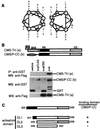
Dimerization of CMS is mediated through the COOH-terminal CC region. (A) Helical wheel representation of the putative leucine zipper in the CC region of CMS. (B) 293T cells were cotransfected with either empty vectors, GST vector, and flag-tagged CMS-TH (a), FLAG vector and GST-tagged CMS/P-CC (b), or CMS-TH and CMS/P-CC. Immunoprecipitates (IP) were analyzed by WB using anti-flag antibody (Upper). The expression levels were analyzed in total cell lysates by WB. (C) Mapping of the dimerization site in CMS. CMS target constructs for expression in yeast were introduced in vector pACT2 and transfected together with the bait CMS/P-CC cloned in pAS2–1. Transformants were grown on plates lacking leucine and tryptophane. Three single colonies were analyzed for interaction in a β-galactosidose filter assay (19). −, no interaction. +, interaction.

References

    1. Keely P, Parise L, Juliano R. Trends Cell Biol. 1998;8:101–106. - PubMed
    1. Clezardin P. Cell Mol Life Sci. 1998;54:541–548. - VSports在线直播 - PMC - PubMed
    1. Clark E A, Brugge J S. Science. 1995;268:233–239. - PubMed
    1. Howe A, Aplin A E, Alahari S K, Juliano R L. Curr Opin Cell Biol. 1998;10:220–231. - PubMed
    1. Ojaniemi M, Vuori K. J Biol Chem. 1997;272:25993–25998. - PubMed

Publication types

MeSH terms (V体育官网入口)

"V体育平台登录" Substances

Associated data (VSports在线直播)

LinkOut - more resources