VSports app下载 - Skip to main page content
U.S. flag

An official website of the United States government

Dot gov

The . gov means it’s official VSports app下载. Federal government websites often end in . gov or . mil. Before sharing sensitive information, make sure you’re on a federal government site. .

Https

The site is secure V体育官网. The https:// ensures that you are connecting to the official website and that any information you provide is encrypted and transmitted securely. .

. 1999 Jun;19(6):4390-404.
doi: 10.1128/MCB.19.6.4390.

Btf, a novel death-promoting transcriptional repressor that interacts with Bcl-2-related proteins

Affiliations

Btf, a novel death-promoting transcriptional repressor that interacts with Bcl-2-related proteins

G M Kasof et al. Mol Cell Biol. 1999 Jun.

Abstract

The adenovirus E1B 19,000-molecular-weight (19K) protein is a potent inhibitor of apoptosis and cooperates with E1A to transform primary rodent cells. E1B 19K shows sequence and functional homology to the mammalian antiapoptotic gene product, Bcl-2. Like Bcl-2, the biochemical mechanism of E1B 19K function includes binding to and antagonization of cellular proapoptotic proteins such as Bax, Bak, and Nbk/Bik. In addition, there is evidence that E1B 19K can affect gene expression, but whether this contributes to its antiapoptotic function has not been determined. In an effort to further understand the functions of E1B 19K, we screened for 19K-associated proteins by the yeast two-hybrid system. A novel protein, Btf (Bcl-2-associated transcription factor), that interacts with E1B 19K as well as with the antiapoptotic family members Bcl-2 and Bcl-xL but not with the proapoptotic protein Bax was identified. btf is a widely expressed gene that encodes a protein with homology to the basic zipper (bZip) and Myb DNA binding domains. Btf binds DNA in vitro and represses transcription in reporter assays. E1B 19K, Bcl-2, and Bcl-xL sequester Btf in the cytoplasm and block its transcriptional repression activity. Expression of Btf also inhibited transformation by E1A with either E1B 19K or mutant p53, suggesting a role in either promotion of apoptosis or cell cycle arrest. Indeed, the sustained overexpression of Btf in HeLa cells induced apoptosis, which was inhibited by E1B 19K. Furthermore, the chromosomal localization of btf (6q22-23) maps to a region that is deleted in some cancers, consistent with a role for Btf in tumor suppression. Thus, btf may represent a novel tumor suppressor gene residing in a unique pathway by which the Bcl-2 family can regulate apoptosis. VSports手机版.


Figures

FIG. 1
FIG. 1
BP-1 interacts with E1B 19K in yeast. (A) The yeast two-hybrid assay was used to demonstrate binding between BP-1 and E1B 19K. Growth in the presence of histidine indicates that both plasmids can be expressed in yeast, and growth in the absence of histidine demonstrates an interaction between the two proteins. Apc-2 represents an irrelevant hydrophobic protein used as a negative control. The specificity of the interaction between BP-1 and E1B 19K was tested by using related proteins (Bcl-2, Bcl-xL, and Bax) as well as missense mutants of E1B 19K (pm7, pm51, pm87, and pm102). (B) The minimal regions of E1B 19K required for interaction with BP-1 were mapped by using a series of deletion mutants. (C) Schematic representation of E1B 19K showing the missense and deletion mutants tested in the two-hybrid assay. Regions I and III indicate the locations of the Bcl-2 homology domains BH1 and BH3, respectively. E1B 19K does not contain recognizable BH2 or BH4 domains.
FIG. 1
FIG. 1
BP-1 interacts with E1B 19K in yeast. (A) The yeast two-hybrid assay was used to demonstrate binding between BP-1 and E1B 19K. Growth in the presence of histidine indicates that both plasmids can be expressed in yeast, and growth in the absence of histidine demonstrates an interaction between the two proteins. Apc-2 represents an irrelevant hydrophobic protein used as a negative control. The specificity of the interaction between BP-1 and E1B 19K was tested by using related proteins (Bcl-2, Bcl-xL, and Bax) as well as missense mutants of E1B 19K (pm7, pm51, pm87, and pm102). (B) The minimal regions of E1B 19K required for interaction with BP-1 were mapped by using a series of deletion mutants. (C) Schematic representation of E1B 19K showing the missense and deletion mutants tested in the two-hybrid assay. Regions I and III indicate the locations of the Bcl-2 homology domains BH1 and BH3, respectively. E1B 19K does not contain recognizable BH2 or BH4 domains.
FIG. 2
FIG. 2
Northern blotting was performed to determine btf expression in various tissues (A) and cancer cell lines (B). Two transcripts were observed, at 5 kb (btfL) and 3 kb (btfS). These transcripts appeared to be ubiquitously expressed, except that btf was not detected in Raji cells derived from Burkitt’s lymphoma. β-Actin expression was examined to assess the quality of the RNA and to control for loading efficiency.
FIG. 3
FIG. 3
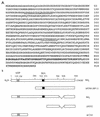
(A) Amino acid sequence of Btf. BtfL, identified in the GenBank database (accession no. D79986), is predicted to encode a 918-amino-acid protein, shown here. BtfS, which was obtained in the λ screen, contains the complete BtfL sequence except that it is missing 49 amino acids between residues 797 and 846 (bold) present in BtfL and BP-1. The underlined regions represent the positions of nuclear localization sequences. (B) A schematic representation of the Btf variants and deletion mutants used to characterize Btf function. The shaded and solid boxes represent the locations of putative bZIP and Myb-DNA binding domains, respectively. BP-1 (ΔN384), identified in the yeast two-hybrid screen for E1B 19K binding proteins, contains residues 384 to 918 of BtfL. Deletion mutants of BtfS, ΔN552, and ΔC210 were used in transcriptional reporter assays to determine the regions of BtfS that contribute to transcriptional repression.
FIG. 4
FIG. 4
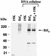
BtfS binds to DNA-cellulose in vitro. [35S]methione-labeled, in vitro-translated Myc-BtfS and E1B 19K were prepared by using the TnT T7 reticulocyte lysate system and incubated with native DNA-cellulose in NETN buffer for 2 h. Samples were washed five times in NETN, and the proteins were resolved by SDS-PAGE (17% polyacrylamide). Immunoprecipitated proteins (lanes 1 and 2) were analyzed to confirm the presence of the in vitro-translated products.
FIG. 5
FIG. 5
In vitro interactions between BtfS and antiapoptotic Bcl-2 family members. [35S]methionine-labeled, in vitro-translated Myc-BtfS or Myc-Bax was combined with V5/His-E1B 19K, Bcl-2, Flag–Bcl-xL, or luciferase prepared by using the TnT T7 reticulocyte lysate system. The proteins were immunoprecipitated with anti-Myc monoclonal antibody in NETN buffer for 1.5 h followed by protein A-Sepharose for 0.5 h. Samples were washed in NETN buffer, resolved by SDS-PAGE (14% polyacrylamide), and visualized by autoradiography. In addition, 1 μl of each of translation reaction mixture was analyzed to verify that equal amounts of proteins were used in the binding assay.
FIG. 6
FIG. 6
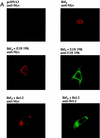
Nuclear subcellular localization of BtfS is altered by E1B 19K, Bcl-2, and BtfL. (A) HeLa cells were transfected with expression plasmid pcDNA3-Myc-BtfS alone or with pCMV E1B 19K or pcDNA3–Bcl-2 as indicated. The cells were fixed 24 h posttransfection and double stained against Myc and E1B 19K or Bcl-2. Magnification, ×1,000. (B) HeLa cells expressing Bcl-xL were transfected with pcDNA3-Myc-BtfS and stained against Myc 24 h posttransfection. Values represent the percentages of nuclear BtfS expression (BtfS/total BtfS) in these cells.
FIG. 6
FIG. 6
Nuclear subcellular localization of BtfS is altered by E1B 19K, Bcl-2, and BtfL. (A) HeLa cells were transfected with expression plasmid pcDNA3-Myc-BtfS alone or with pCMV E1B 19K or pcDNA3–Bcl-2 as indicated. The cells were fixed 24 h posttransfection and double stained against Myc and E1B 19K or Bcl-2. Magnification, ×1,000. (B) HeLa cells expressing Bcl-xL were transfected with pcDNA3-Myc-BtfS and stained against Myc 24 h posttransfection. Values represent the percentages of nuclear BtfS expression (BtfS/total BtfS) in these cells.
FIG. 7
FIG. 7
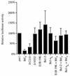
Reporter assay demonstrating that BtfS is a transcription repressor and is inhibited by Bcl-2-like proteins. HeLa cells were transfected with 2.5 μg of the reporter construct (luciferase construct containing GAL4 DNA binding sites within its promoter), 2.5 μg of GAL4 DNA binding domain fusion genes (pm1-BtfS, pm1-ΔN552, and pm1-ΔC210 or empty pm1 vector control), and 2.5 μg of bcl-2 family gene (pCMV-E1B 19K, pcDNA3–Bcl-2, pcDNA3-Flag–Bcl-xL, or empty pcDNA3 vector control). The cells were harvested 24 hours posttransfection, and the luciferase activity was measured in a scintillation counter with the luciferase substrate luciferin. Values were normalized for protein concentrations measured by the Bradford assay and graphed as a percentage of the result for the negative control (empty pm1 vector). The experiment was performed six times, and the high and low values for each sample were dropped. Bars indicate standard deviations (n = 4).
FIG. 8
FIG. 8
BtfS inhibits transformation by E1A and E1B 19K (A) or p53DD (B). Primary BRK cells were transfected with carrier DNA along with a linearized test DNA (15 μg of pCMV-E1A, 15 μg of pCMV-E1B 19K or pCMV-p53DD, and 45 μg of pcDNA3-Myc-BtfS). DNA concentrations were kept constant by using appropriate empty vectors. The cells were cultured for 3 to 4 weeks and then stained with Giemsa. Foci were counted from four dishes per condition. Bars indicate standard deviations (n = 4).
FIG. 9
FIG. 9
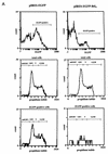
BtfS-mediated cell death occurs by apoptosis. (A) Representative FACS scan from Table 1 at 72 h after transfection of pIRES-EGFP-BtfS and the empty pIRES-EGFP vector into HeLa cells. Cell cycle kinetics from propidium iodide staining are shown for the total cell population as well as the EGFP-positive cells. (B) Transfected cells were incubated with Hoechst dye 72 h posttransfection to visualize the DNA. Cells transfected with pIRES-EGFP-BtfS (right) were compared to those transfected with control pIRES-EGFP (left). Arrows for each set correspond to the same cell visualized with bright field (top), EGFP (green; middle), and Hoechst dye (blue; bottom). The presence of condensed chromatin in the presence of BtfS indicates cell death by apoptosis. Original magnification, ×1,000.
FIG. 9
FIG. 9
BtfS-mediated cell death occurs by apoptosis. (A) Representative FACS scan from Table 1 at 72 h after transfection of pIRES-EGFP-BtfS and the empty pIRES-EGFP vector into HeLa cells. Cell cycle kinetics from propidium iodide staining are shown for the total cell population as well as the EGFP-positive cells. (B) Transfected cells were incubated with Hoechst dye 72 h posttransfection to visualize the DNA. Cells transfected with pIRES-EGFP-BtfS (right) were compared to those transfected with control pIRES-EGFP (left). Arrows for each set correspond to the same cell visualized with bright field (top), EGFP (green; middle), and Hoechst dye (blue; bottom). The presence of condensed chromatin in the presence of BtfS indicates cell death by apoptosis. Original magnification, ×1,000.

References

    1. Antonsson B, Conti F, Ciavatta A, Montessuit S, Lewis S, Martinou I, Bernasconi L, Bernard A, Mermod J-J, Mazzei G, Maundrell K, Gambale F, Sadoul R, Martinou J-C. Inhibition of Bax channel-forming activity by Bcl-2. Science. 1997;277:370–372. - "V体育平台登录" PubMed
    1. Barry M A, Behnke C A, Eastman A. Activation of programmed cell death (apoptosis) by cisplatin, other anticancer drugs, toxins and hyperthermia. Biochem Pharmacol. 1990;40:2353–2362. - PubMed
    1. Bazer L S, Deeg H J. Ultraviolet B-induced DNA fragmentation (apoptosis) in activated T-lymphocytes and Jurkat cells is augmented by inhibition of RNA and protein synthesis. Exp Hematol. 1992;20:80–86. - PubMed
    1. Beg A A, Baltimore D. An essential role for NF-κB in preventing TNF-α-induced cell death. Science. 1996;274:782–784. - "VSports" PubMed
    1. Boise L H, Gonzalez-Garcia M, Postema C E, Ding L, Lindsten T, Turka L A, Mao X, Nuñez G, Thompson C. bcl-x, a bcl-2-related gene that functions as a dominant regulator of apoptotic death. Cell. 1993;74:597–608. - PubMed

Publication types

VSports app下载 - MeSH terms

Substances (VSports手机版)